42 label the image of a compound light microscope
Parts of a Compound Microscope and Their Functions This number represents the compound microscope magnification power of the device. With the eyepiece, you may see a magnified image of the thing. Mirror: It is either fastened to the pillar or the lower end of the arm. On one side, it has a flat mirror, while on the other, it has a concave mirror. Compound Microscope Parts, Function, & Diagram | What is a Compound ... The body of the compound light microscope is the main part of the microscope, not to include the lights, focusing block, or stand of the microscope. The objective lenses and eyepiece are a part of ...
PDF COMPOUND LIGHT MICROSCOPE LAB - Springfield Public Schools ( 3.(Describe(what(happens(to(the(letter("e"(in(the(viewing(area(when(you(move(ittotheright?Totheleft? Up?Down?(((C.(Using(the(prepared(slide(provided(by(your ...
Label the image of a compound light microscope
Bright-field microscope (Compound light microscope) - Diagram (Parts ... Bright-field Microscope. A bright-field microscope, also known as a compound light microscope is among the simplest of optical microscopes. Optical microscopes employ visible light and a series of lenses to magnify the specimen and view it in detail. A bright-field microscope uses light rays to create a dark image against a bright background ... Compound Microscope - Diagram (Parts labelled), Principle and Uses Using a combination of lenses, the working principle of a compound microscope is that a highly magnified image of the specimen is formed at the least possible distance from the distinct vision of an eye that is held very close to the eyepiece of the microscope when the specimen is placed just beyond the focus of the objective lens. microscopeinternational.com › fluorescence-microscopyFluorescence Microscopy - New York Microscope Company Dec 16, 2020 · A fluorescence microscope uses a higher intensity light to illuminate the samples. Parts of a Fluorescence Microscope A powerful light source (xenon or mercury arch lamp) : The light emitted from the mercury arc lamp is 10-100 times brighter than most incandescent lamps and provides light in a wide range of wavelengths, from ultra-violet to the ...
Label the image of a compound light microscope. Light Microscope Definitions..jpg - MICROSCOPE IDENTIFICATION Using the ... Arm: Supports the tube and connects it to the base 3. Objective lens: Several lenses combined to magnify and project a larger image. 4. Coarse focus adjustment tho : Moves the objetive lens up and down. 5. Fine focus adjustment knob: Sharpen the focus quality 6. Base with light source: FIluminator of the microscope 7. Nosepiece: Holds the ... Compound Light Microscope Optics, Magnification and Uses Magnification. In order to ascertain the total magnification when viewing an image with a compound light microscope, take the power of the objective lens which is at 4x, 10x or 40x and multiply it by the power of the eyepiece which is typically 10x. Therefore, a 10x eyepiece used with a 40X objective lens, will produce a magnification of 400X. Labelled Diagram of Compound Microscope - Biology Discussion The below mentioned article provides a labelled diagram of compound microscope. Part # 1. The Stand: The stand is made up of a heavy foot which carries a curved inclinable limb or arm bearing the body tube. The foot is generally horse shoe-shaped structure (Fig. 2) which rests on table top or any other surface on which the microscope in kept. www2.nau.edu › lrm22 › lessonsMicroscope Notes - Northern Arizona University Microscope Drawings. When drawing what you see under the microscope, follow the format shown below. It is important to include a figure label and a subject title above the image. The species name (and common name if there is one) and the magnification at which you were viewing the object should be written below the image.
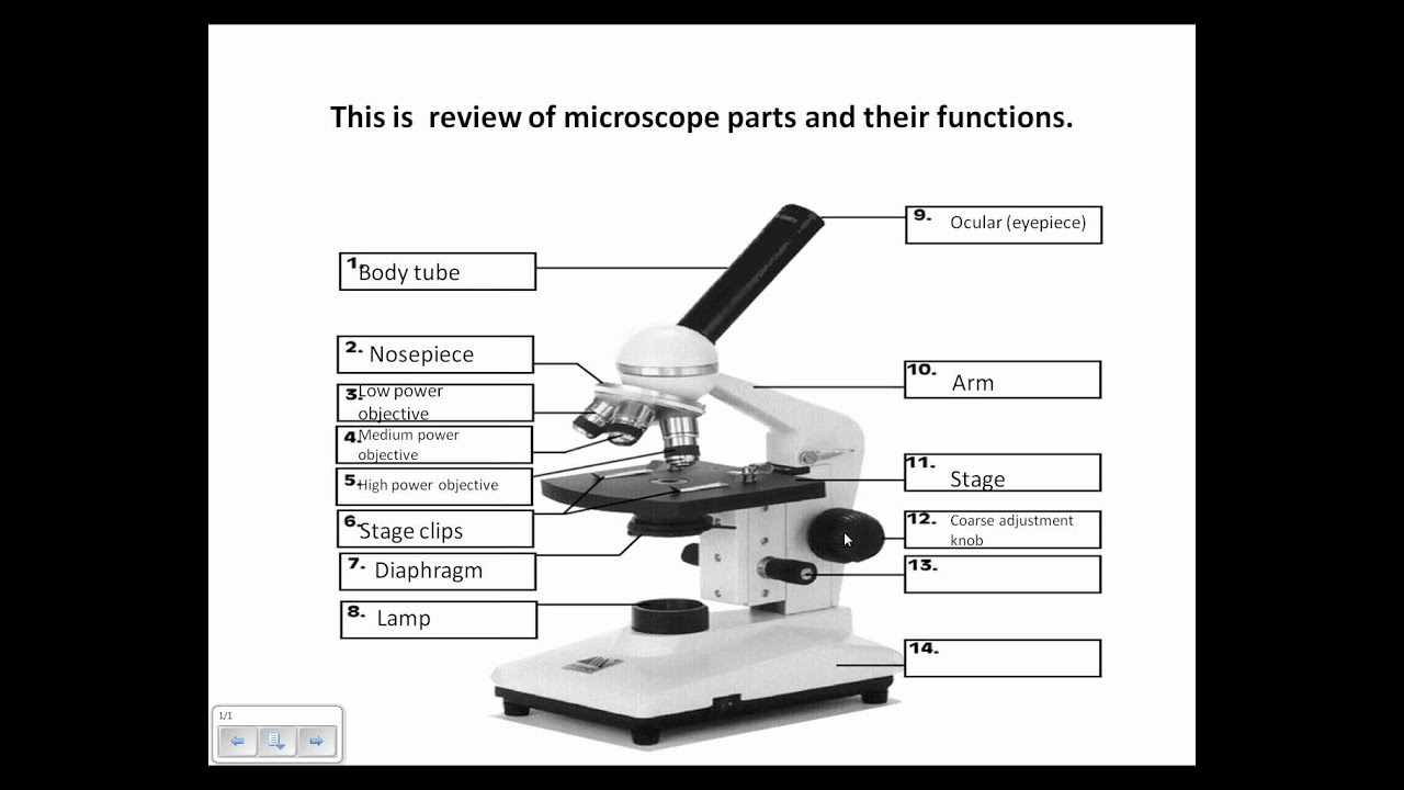
Compound Light Microscope: Everything You Need to Know A compound light microscope is a type of light microscope that uses a compound lens system, meaning, it operates through two sets of lenses to magnify the image of a specimen. It's an upright microscope that produces a two-dimensional image and has a higher magnification than a stereoscopic microscope. PDF Parts of the Light Microscope - Science Spot to SHARPEN the image G. BASE Supports the MICROSCOPE D. STAGE CLIPS HOLD the slide in place C. OBJECTIVE LENSES Magnification ranges from 10 X to 40 X F. LIGHT SOURCE Projects light UPWARDS through the diaphragm, the SPECIMEN, and the LENSES H. DIAPHRAGM Regulates the amount of LIGHT on the specimen E. STAGE Supports the SLIDE being viewed K. ARM Compound Microscope Parts - Labeled Diagram and their Functions - Rs ... A compound microscope is the most common type of light (optical) microscopes. The term "compound" refers to the microscope having more than one lens. Basically, compound microscopes generate magnified images through an aligned pair of the objective lens and the ocular lens. compound microscope parts (labeling) Flashcards | Quizlet Start studying compound microscope parts (labeling). Learn vocabulary, terms, and more with flashcards, games, and other study tools. ... light source of the microscope. what is 8? eyepiece (ocular lens) - magnifying piece that is looked into in order to see the specimen ... knob that brings the image to a sharper focus. what is 13? base - the ...
› game › kVf84IlEU34Sheep Brain Quiz - PurposeGames.com Compound Light Microscope 17p Shape Quiz. ... Label the Skeleton 25p Image Quiz. ... Label the Integumentary System 10p Image Quiz. PurposeGames Create. Play. 16 Parts of a Compound Microscope: Diagrams and Video In compound microscopes with two eye pieces there are prisms contained in the body that will also split the beam of light to enable you to view the image through both eye pieces. 2. Arm. The arm of the microscope is another structural piece. The arm connects the base of the microscope to the head/body of the microscope. PDF The Compound Light Microscope The Compound Light Microscope TASK Refer to page 605 in your text to: 1. Name each of the structures described in the table to the right. 2. Match each structure to the letter in the diagram below. ** ALWAYS USE TWO HANDS TO CARRY A MICROSCOPE** Letter Structure Function joins body tube to base supports the entire microscope Label the image of a compound light microscope - Soetrust Which was the first cell viewed by the light microscope? Which of the following is true regarding the properties of… The compound below is treated with n-bromosuccinimide (nbs)…
Molecular Expressions Microscopy Primer: Specialized Microscopy ...
Parts of A Compound Microscope » Microscope Club A compound microscope is a type of microscope that has compound lenses, and operates through light microscopy techniques. It is a high magnification and high power microscope suitable for viewing minute details of small specimens that are invisible to the naked eye.
Principles Of Biology Lab Quiz - ProProfs Quiz
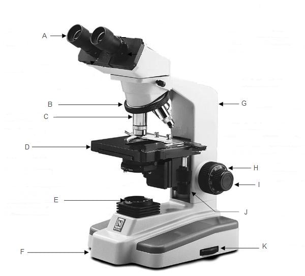
Solved Microscope parts/labeling 9 Label the image of a - Chegg question: microscope parts/labeling 9 label the image of a compound light microscope using the terms provided. 1 points eyepiece eyepiece light source references references base arm slide holder arm stage mechanical stage fine adjustment knob power switch objectives mindeneer microscopy lab homework saved help save & exit submit eyepiece light …
Microscope Review.wmv - YouTube
Compound Microscope: Parts of Compound Microscope - BYJUS A compound microscope is an intricate gathering of a combination of lenses that renders a highly maximized and magnified image of microscopic living entities and other complex details or tissues and cells. Diagram Parts of the Compound Microscope Parts Of Compound Microscope The parts of the compound microscope can be categorized into:

Compound microscope clipart 5 » Clipart Station
Label the above components of the compound light microscope A Ocular ... Label the above components of the compound light 4. Label the above components of the compound light microscope: A. Ocular lens (eyepiece) F. Rheostat B. Stage controls G. Spring loaded stage clip C. Coarse focus H. Illuminator D. Fine focus I. Iris diaphragm E. Objective lens 5.
23 Label And Color The Parts Of Both Microscopes - Labels 2021
Microscope Parts and Functions Microscope Parts and Functions With Labeled Diagram and Functions How does a Compound Microscope Work?. Before exploring microscope parts and functions, you should probably understand that the compound light microscope is more complicated than just a microscope with more than one lens.. First, the purpose of a microscope is to magnify a small object or to magnify the fine details of a larger ...

Bio: Lab 2 The Microscope Flashcards by ProProfs
› articles › s41396/021/01078-7Light-driven carbon dioxide reduction to methane by ... - Nature Aug 02, 2021 · The direct conversion of CO2 to value-added chemical commodities, thereby storing solar energy, offers a promising option for alleviating both the current energy crisis and global warming.

Microscope parts and functions
Compound Microscope: Definition, Diagram, Parts, Uses, Working ... - BYJUS The objective lens is a compound lens that forms a real inverted image of the image inside the body tube. Ocular Lens The ocular lens is also known as the eyepiece. The image of microscopic objects can be viewed through these lenses. There are four types of magnification that can take place in the ocular lens: 5X 10X 15X 20X

The Compound Microscope
The Compound Light Microscope Label the following parts on ... The Compound Light Microscope Label the following parts on the diagram: ... E. moves the body tube freely up and down to focus the image. F. provides light ...2 pages
Post a Comment for "42 label the image of a compound light microscope"