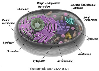
45 cell diagram with labels
Cell diagram with labels - Graph Diagram Cell Diagram With Labels This human anatomy diagram with labels depicts and explains the details and or parts of the Cell Diagram With Labels. Human anatomy diagrams and charts show internal organs, body systems, cells, conditions, sickness and symptoms information and/or tips to ensure one lives in good health. Skin Diagram with Detailed Illustrations and Clear Labels Skin Diagram. The largest organ in the human body is the skin, covering a total area of about 1.8 square meters. The skin is tasked with protecting our body from the external elements as well as microbes. Interesting Note: The skin is also responsible for maintaining our body temperature – this was apparent in victims who were subjected to the medival torture of being skinned alive. …
Cell Diagrams - The Biology Corner Open Google Draw and import the diagram. Then use "insert" to create text boxes where students can fill in the labels. Don't forget when assigning this to students on Google classroom to make a copy for each student. You can leave documents in an uneditable form and students can use an addon like "Kami" to annotate the document.

Cell diagram with labels
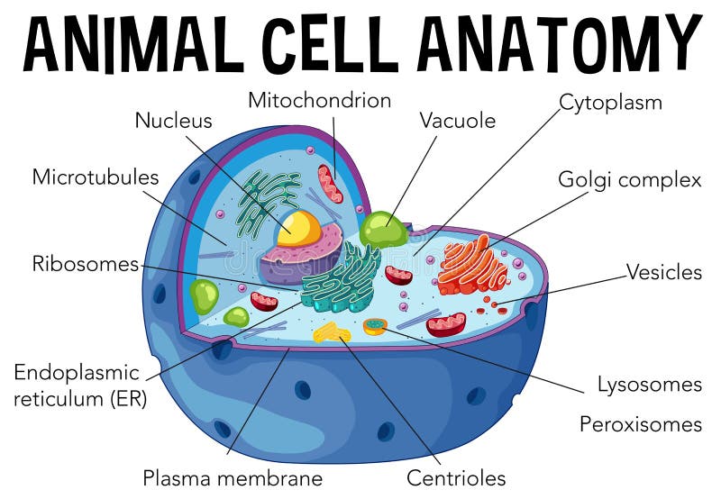
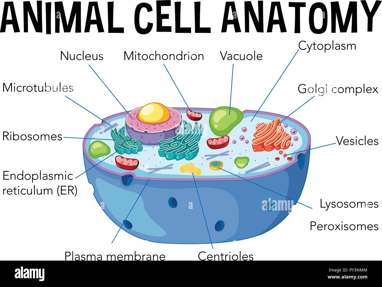
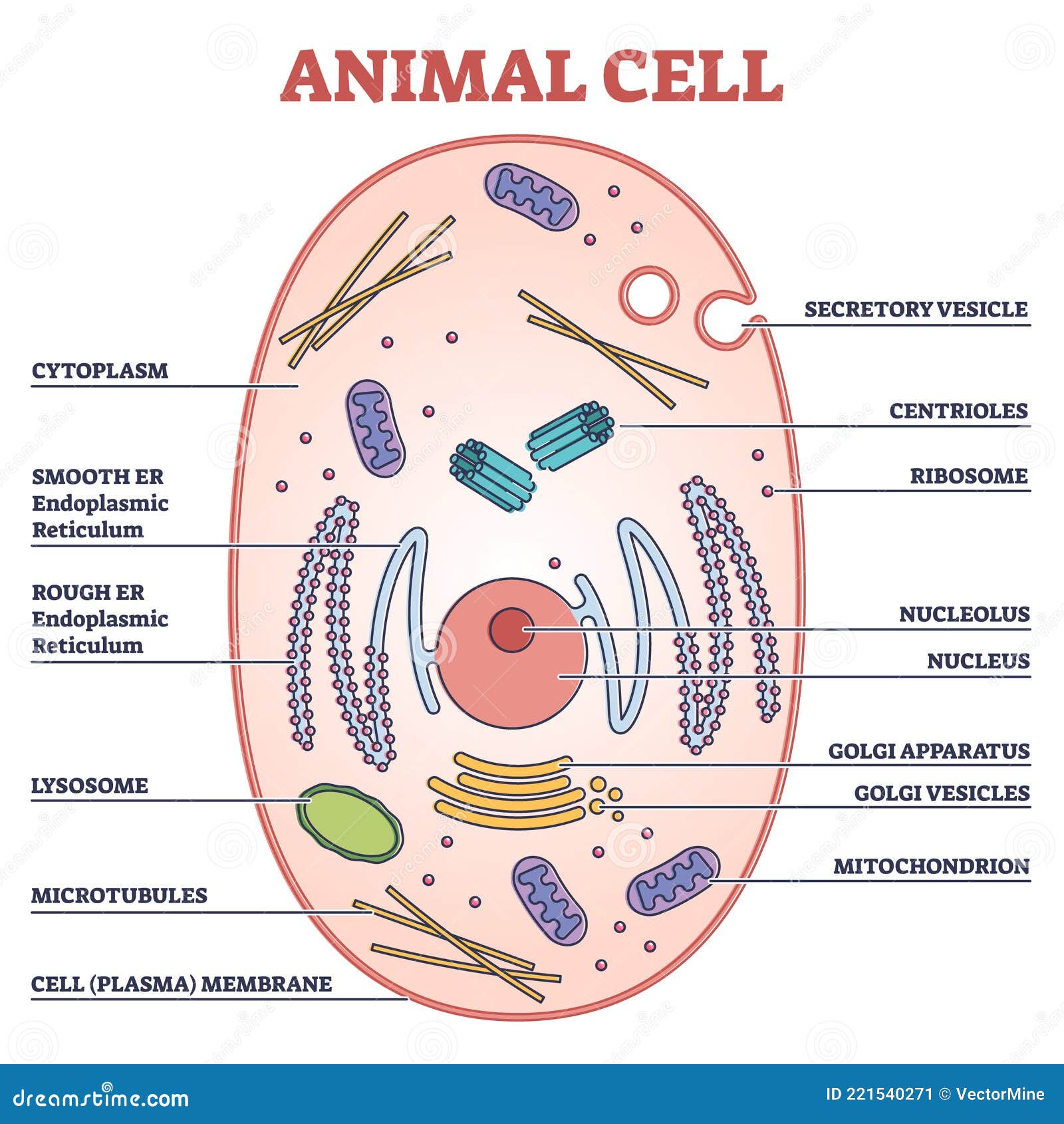
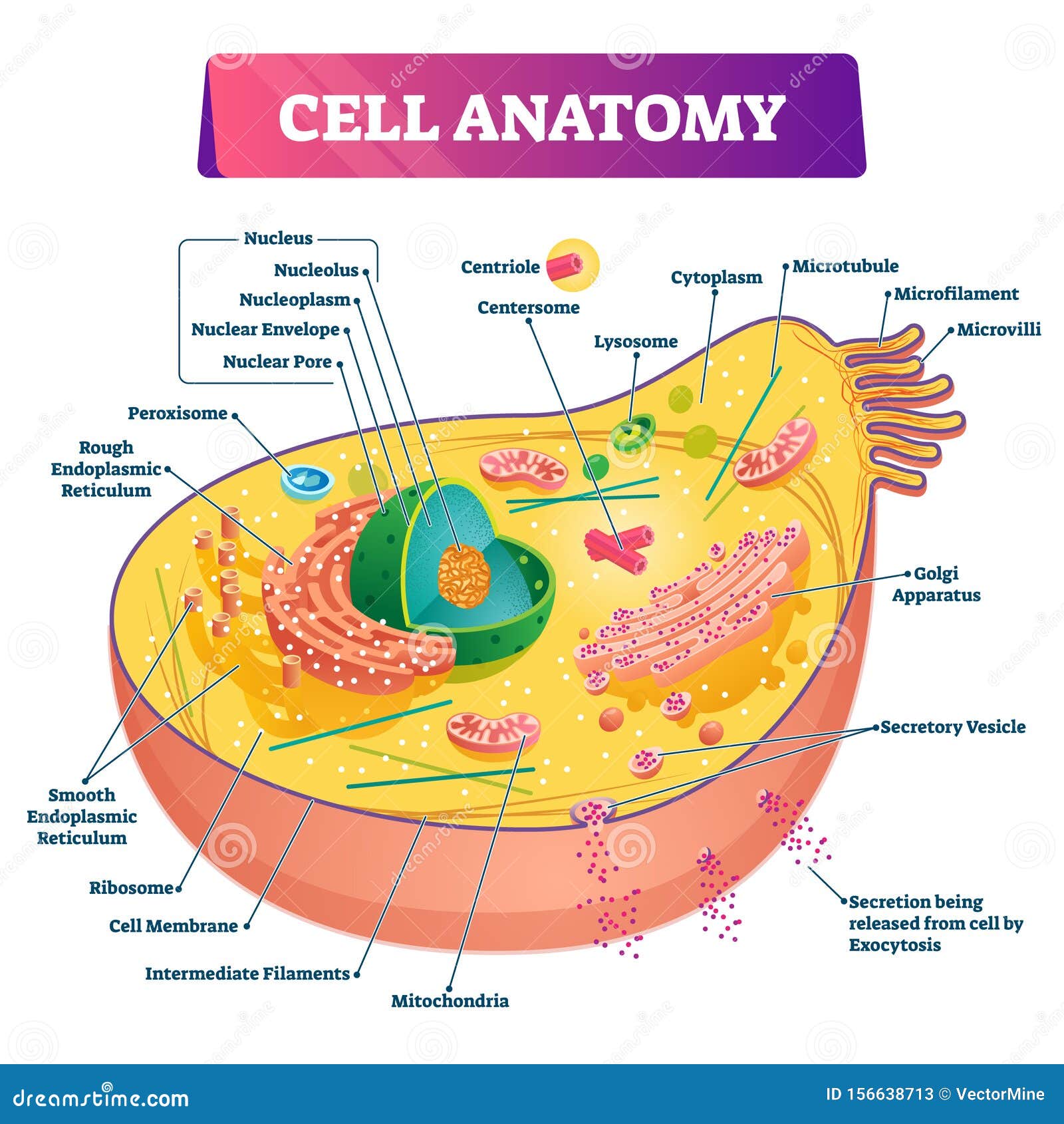
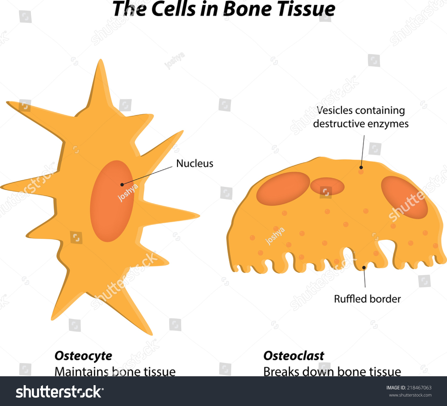
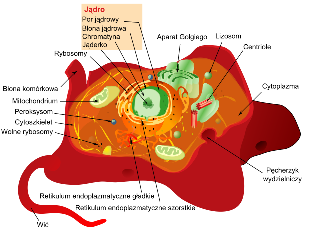
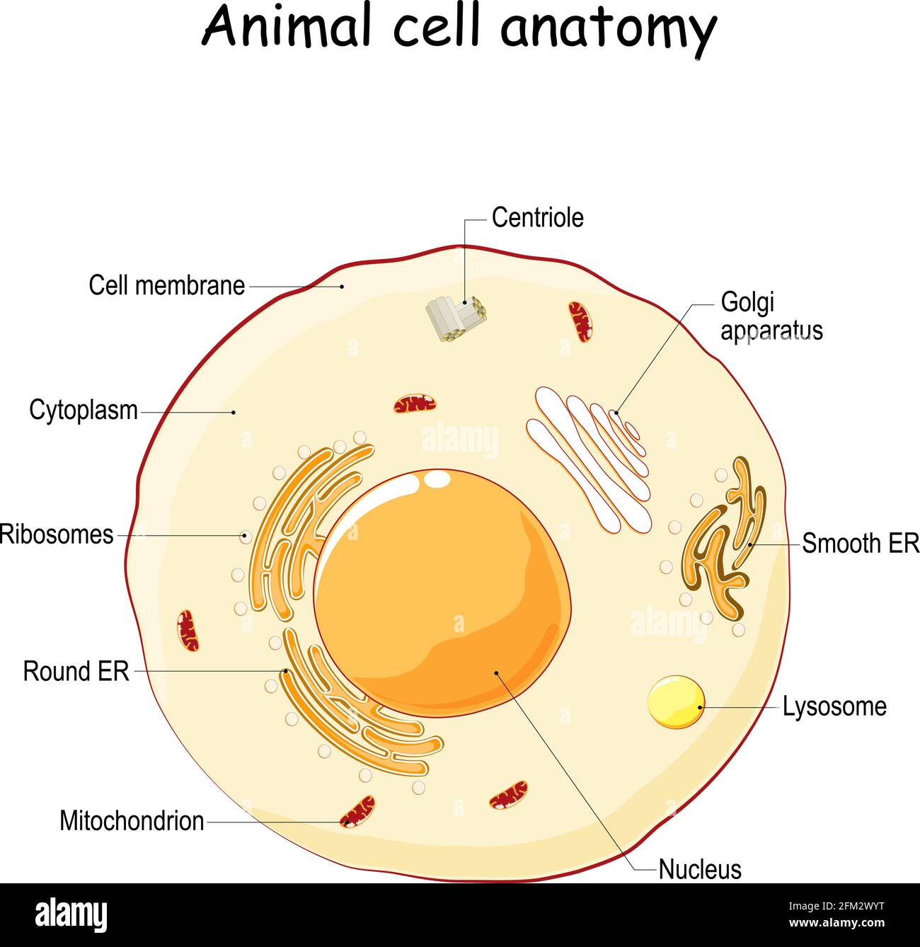
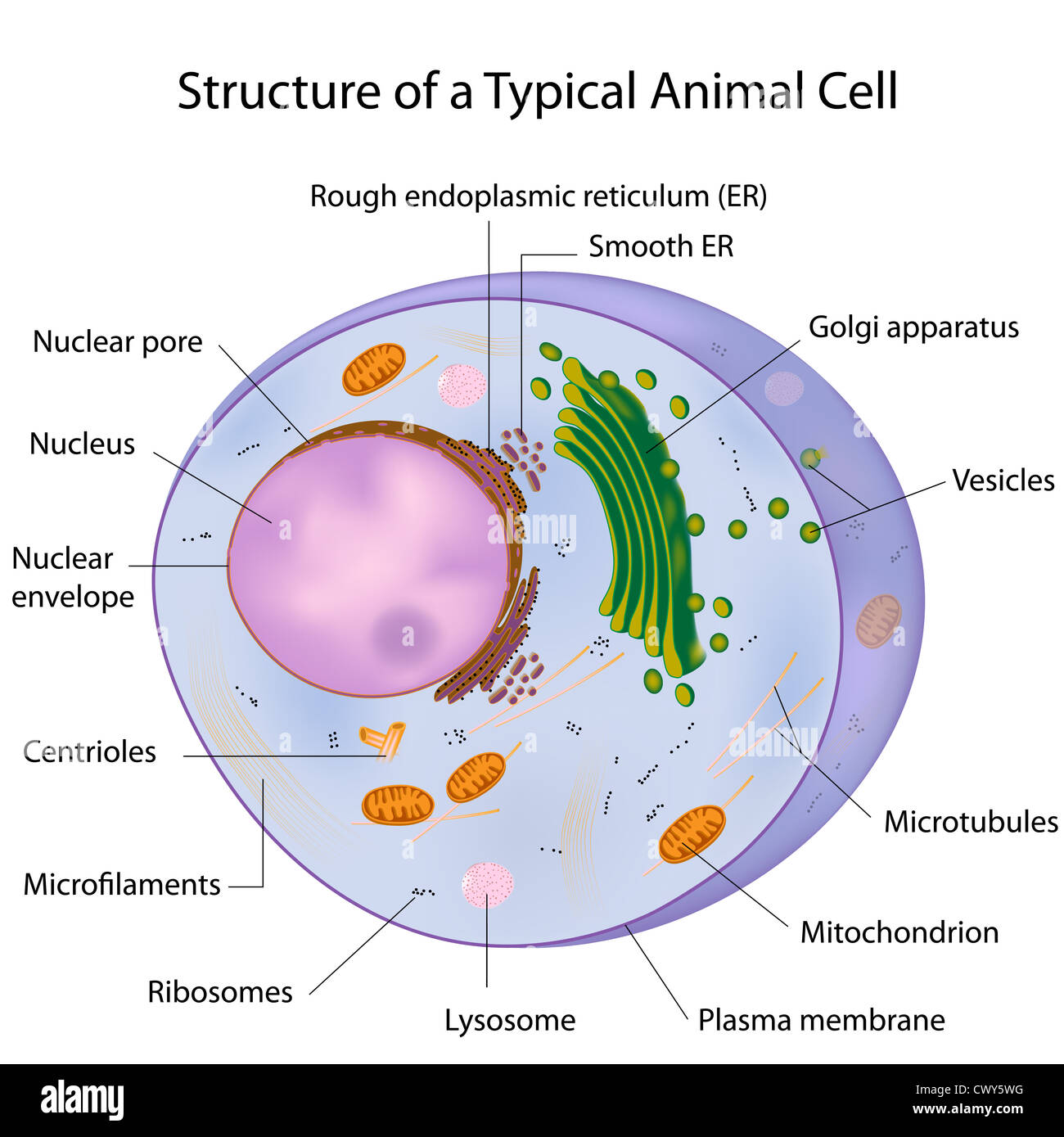
A Well-labelled Diagram Of Animal Cell With Explanation The animal cell diagram is widely asked in Class 10 and 12 examinations and is beneficial to understand the structure and functions of an animal. A brief explanation of the different parts of an animal cell along with a well-labelled diagram is mentioned below for reference. Also Read Different between Plant Cell and Animal Cell Plant Cells: Labelled Diagram, Definitions, and Structure Plastids and Chloroplasts. Plants make their own food through photosynthesis. Plant cells have plastids, which animal cells don't. Plastids are organelles used to make and store needed compounds. Chloroplasts are the most important of plastids. They convert light energy from the sun into sugar and oxygen. The most exposed parts of the plants ... Human Cell Diagram, Parts, Pictures, Structure and Functions Diagram of the human cell illustrating the different parts of the cell. Cell Membrane. The cell membrane is the outer coating of the cell and contains the cytoplasm, substances within it and the organelle. It is a double-layered membrane composed of proteins and lipids. The lipid molecules on the outer and inner part (lipid bilayer) allow it to ...
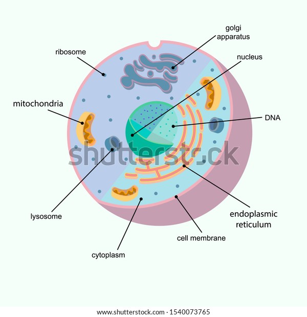
Cell diagram with labels. Red Blood Cell Diagram Labeled stock illustrations Browse 19 red blood cell diagram labeled stock illustrations and vector graphics available royalty-free, or start a new search to explore more great stock images and vector art. Newest results. Human gas exchange system vector illustration. Oxygen travel from lungs to heart, to all body cells and back to lungs as CO2. Animal Cells: Labelled Diagram, Definitions, and Structure The endoplasmic reticulum (s) are organelles that create a network of membranes that transport substances around the cell. They have phospholipid bilayers. There are two types of ER: the rough ER, and the smooth ER. The rough endoplasmic reticulum is rough because it has ribosomes (which is explained below) attached to it. Chord diagram – from Data to Viz A chord diagram represents flows or connections between several entities (called nodes).Each entity is represented by a fragment on the outer part of the circular layout.Then, arcs are drawn between each entities. The size of the arc is proportional to the importance of the flow. Here is an example displaying the number of people migrating from one country to another. Animal Cell Diagram with Label and Explanation: Cell Structure, Functions Diagram of Animal Cell Below is the diagram of the animal cell which shows the organelles present in it. The cell is covered with cytoplasm which consists of cell organelles in it. The nucleus is covered with a rough Endoplasmic Reticulum and other organelles each designed for a specific purpose.
Label the cell diagram - Teaching resources - Wordwall Label the cell diagram Examples from our community 10000+ results for 'label the cell diagram' Label the Plot Diagram Labelled diagram by Mrsjohnsonncvps G11 Label the Cell Membrane Labelled diagram by Renatathomas Label the Cell Membrane Labelled diagram by Ancclark Label the Plant Cell Labelled diagram by Armstrem G7 Science Identifying Medical Diagnoses and Treatable Diseases by Image ... - Cell 22.02.2018 · (B) Workflow diagram showing overall experimental design describing the flow of optical coherence tomography (OCT) images through the labeling and grading process followed by creation of the transfer learning model, which then underwent training and subsequent testing. The training dataset only included images that passed sufficient quality and diagnostic … CELL MEMBRANE LABEL Diagram | Quizlet Practice labeling the parts of the cell membrane Terms in this set (6) Channel Protein hole or tunnel that particles may pass through to go in / out of cell Marker protein identifies or labels the cell Receptor protein receives information Heads part of the phospholipid that loves water (hydrophili) - points to the most outside and inside of cell How to add Axis Labels (X & Y) in Excel & Google Sheets Make sure the Axis Labels are clear, concise, and easy to understand. Dynamic Axis Titles. To make your Axis titles dynamic, enter a formula for your chart title. Click on the Axis Title you want to change; In the Formula Bar, put in the formula for the cell you want to reference (In this case, we want the axis title “Revenue” in Cell C2”).
Animal Cell Labeled Diagram Pictures, Images and Stock Photos Browse 19 animal cell labeled diagram stock photos and images available, or start a new search to explore more stock photos and images. Newest results. Diagrams of animal and plant cells. Labelled diagrams of typical animal and plant cells with editable layers. Animal cells and plant cells - Cells to systems - BBC Bitesize Part Function Found in; Cell membrane: Controls the movement of substances into and out of the cell: Plant and animal cells: Cytoplasm: Jelly-like substance, where chemical reactions happen PDF Human Cell Diagram, Parts, Pictures, Structure and Functions Diagram of the human cell illustrating the different parts of the cell. Cell Membrane The cell membraneis the outer coating of the cell and contains the cytoplasm, substances within it and the organelle. It is a double-layered membrane composed of proteins and lipids. Cell: Structure and Functions (With Diagram) - Biology Discussion 1. Eukaryotes are sophisticated cells with a well defined nucleus and cell organelles. 2. The cells are comparatively larger in size (10-100 μm). 3. Unicellular to multicellular in nature and evolved ~1 billion years ago. 4. The cell membrane is semipermeable and flexible. 5.
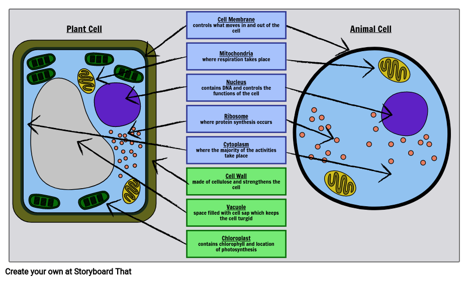
Label Cell Parts | Plant & Animal Cell Activity | StoryboardThat Create a cell diagram with each part of plant and animal cells labeled. Include descriptions of what each organelle does. Click "Start Assignment". Find diagrams of a plant and an animal cell in the Science tab. Using arrows and Textables, label each part of the cell and describe its function.
Liver Diagram with Detailed Illustrations and Clear Labels Liver Diagram. The liver is one of the most important organs in the human body. Anatomically, the liver is a meaty organ that consists of two large sections called the right and the left lobe. The rib cage partly protects the liver and cannot be felt if you were to touch it. However, it can be felt ascending and descending if you were to take a deep breath. The liver weighs an average of …
A Labeled Diagram of the Animal Cell and its Organelles As observed in the labeled animal cell diagram, the cell membrane forms the confining factor of the cell, that is it envelopes the cell constituents together and gives the cell its shape, form, and existence. Cell membrane is made up of lipids and proteins and forms a barrier between the extracellular liquid bathing all cells on the exterior ...
Cell Diagram | Free Cell Diagram Templates - Edrawsoft A free customizable cells diagram template is provided to download and print. Quickly get a head-start when creating your own cell diagram. Here is a simple cell diagram example created by Science Diagram Maker, which is available in different formats. Lab Apparatus List. 64703. 211. Plant Cell Diagram. 19550. 173. Heart Diagram.
03 Label the Cell Diagram | Quizlet Cell Biology 03 Label the Cell STUDY Learn Flashcards Write Spell Test PLAY Match Gravity Created by muskopf1TEACHER Terms in this set (14) Nucleus Control center of the cell Nucleolus Ribosome synthesis Rough Endoplasmic Reticulum Protein transport Smooth Endoplasmic Reticulum Lipid synthesis Mitochondrion Cellular Respiratoin Golgi Apparatus
Plant Cell: Diagram, Types and Functions - Embibe Exams Q.2. How to make a model of a plant cell diagram step by step procedure? Ans: The plant cell diagram can be checked above and on a similar pattern the diagram can be created. Q.3. Why do plant cells possess large-sized vacuoles? Ans: Vacuole functions in the storage of substances, maintenance of osmolarity and sustaining turgor pressure. Q.4.
Interactive Cell Cycle - CELLS alive INTERPHASE. Gap 0. Gap 1. S Phase. Gap 2. MITOSIS . ^ Cell Cycle Overview Cell Cycle Mitosis > Meiosis > Get the Cell Division PowerPoints
Label that Diagram - Cells - Apps on Google Play There are 5 cells presented: Animal Cell, Plant Cell, Amoeba, Paramecium, and Euglena. The player can study the labeled diagrams or play the game of labeling the diagrams. When the game is played, the labels appear in a random order one at a time and the player must tap on the correct dot on the diagram.
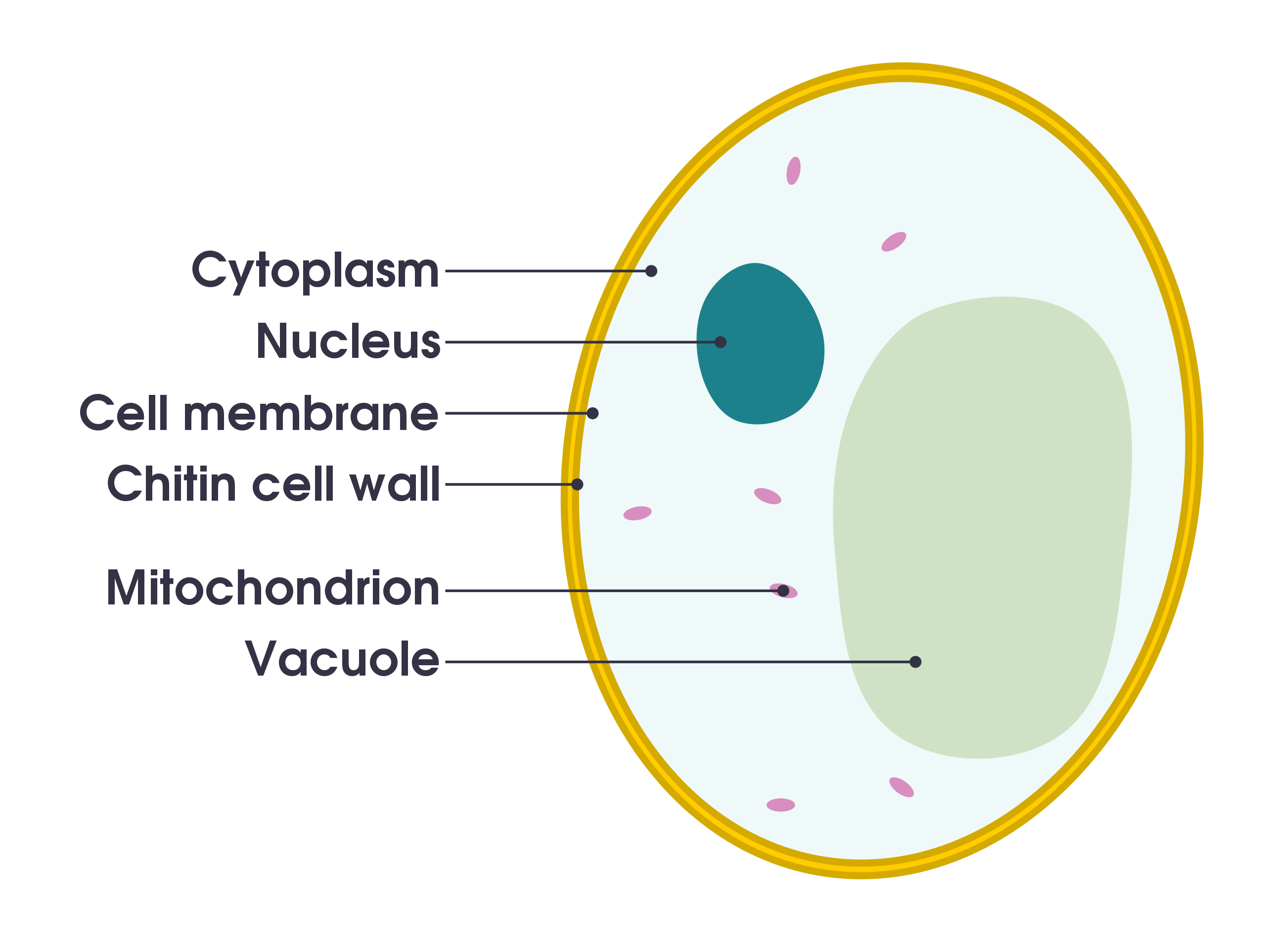
Cell Organelles- Definition, Structure, Functions, Diagram In a plant cell, the cell wall is made up of cellulose, hemicellulose, and proteins while in a fungal cell, it is composed of chitin. A cell wall is multilayered with a middle lamina, a primary cell wall, and a secondary cell wall. The middle lamina contains polysaccharides that provide adhesion and allow binding of the cells to one another.
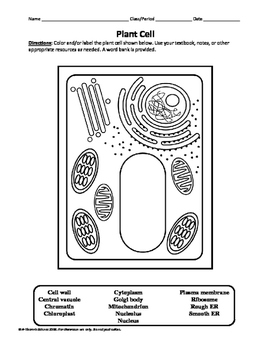
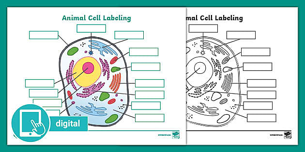
Learn the parts of a cell with diagrams and cell quizzes - Kenhub For this exercise we'll start with an image of a cell diagram ready labeled. Study this and make sure that you're clear about which structure is found where. Cell diagram unlabeled It's time to label the cell yourself! As you fill in the cell structure worksheet, remember the functions of each part of the cell that you learned in the video.
quiver: a modern commutative diagram editor Welcome. quiver is a modern, graphical editor for commutative and pasting diagrams, capable of rendering high-quality diagrams for screen viewing, and exporting to LaTeX via tikz-cd.. quiver is intended to be intuitive to use and easy to pick up. Here are a few tips to help you get started: Click and drag to create new arrows: the source and target objects will be created automatically.
animal and plant cell diagram to label - TeachersPayTeachers Plant and Animal Cell Labeling Diagrams by A-Thom-ic Science 12 $2.00 PDF Three versions of the plant cell worksheet and three versions of the animal cell worksheet allow students of different grade levels and/or skill levels to label and review the parts of each cell type. Can be used as homework or a quiz.
Cells Diagram | Science Illustration Solutions - Edrawsoft Cells Diagram Symbols Edraw software offers you lots of symbols used in cells diagram like cell structure, paramecium, squamous cell, cell division, bacteria, cell membrane, eggs, sperm, zygote, an animal cell, SARS, tobacco mosaic, adenovirus, coliphage, herpesvirus, AIDS, pollen, plant cell model, onion tissue, etc. Cells Diagram Examples
Galvanic Cell: Definition, Diagram and Working - Science ABC 17.01.2022 · Galvanic Cell Diagram. Now, consider this apparatus, which represents a galvanic cell. The first beaker contains zinc sulfate (ZnSO 4) into which a strip of zinc is dipped, while the adjacent beaker contains copper sulfate (CuSO 4) into which a strip of copper is dipped. However, the two strips are connected by an external circuit, a conductor, which is connected to a bulb. …
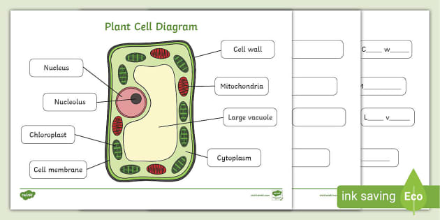
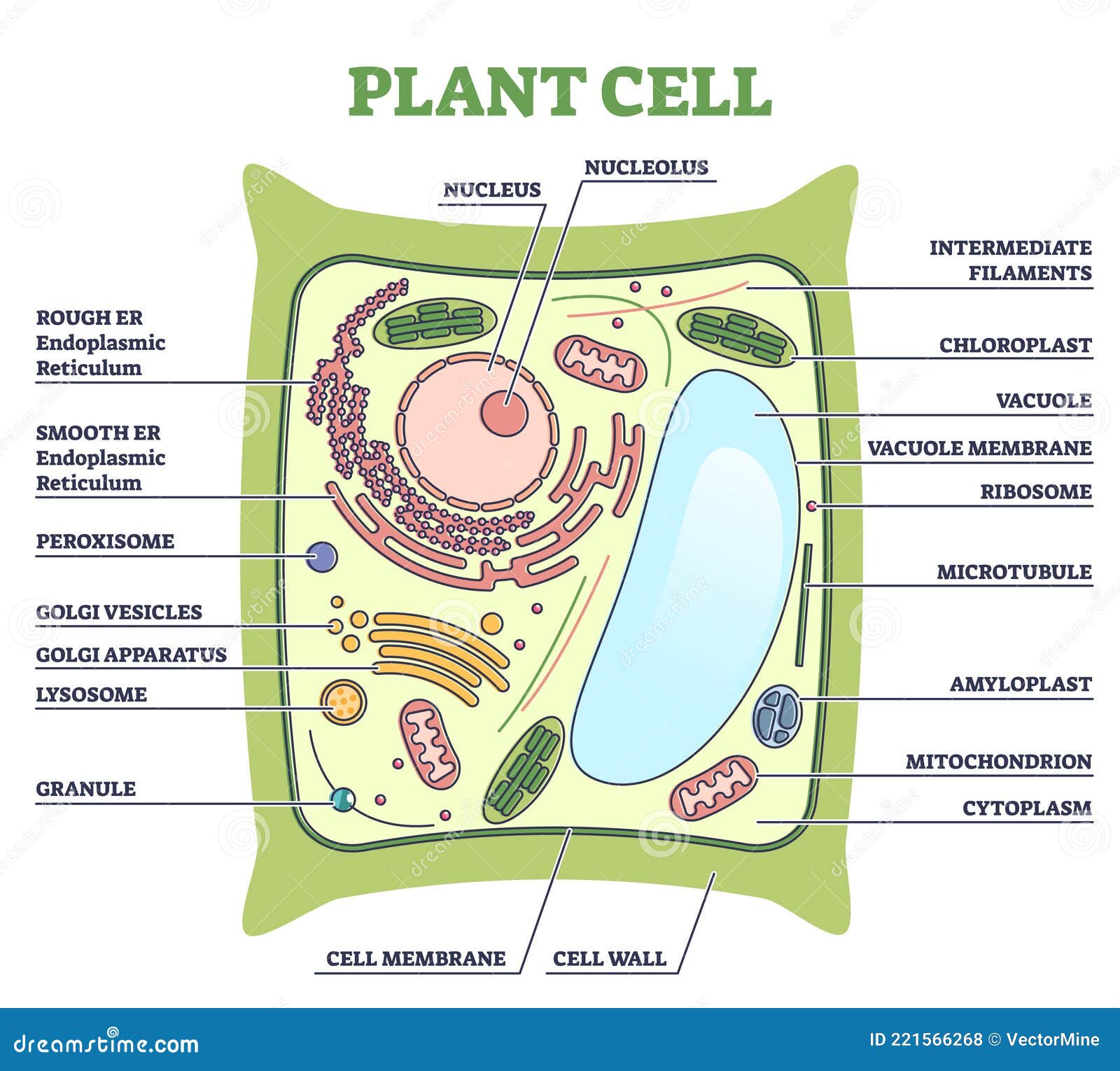
Labeled Plant Cell With Diagrams | Science Trends The parts of a plant cell include the cell wall, the cell membrane, the cytoskeleton or cytoplasm, the nucleus, the Golgi body, the mitochondria, the peroxisome's, the vacuoles, ribosomes, and the endoplasmic reticulum. Parts Of A Plant Cell The Cell Wall Let's start from the outside and work our way inwards.
How to plot a ternary diagram in Excel - Chemostratigraphy.com 13.02.2022 · Ternary diagrams are common in chemistry and geosciences to display the relationship of three variables.Here is an easy step-by-step guide on how to plot a ternary diagram in Excel. Although ternary diagrams or charts are not standard in Microsoft® Excel, there are, however, templates and Excel add-ons available to download from the internet.
A Labeled Diagram of the Plant Cell and Functions of its Organelles A Labeled Diagram of the Plant Cell and Functions of its Organelles We are aware that all life stems from a single cell, and that the cell is the most basic unit of all living organisms. The cell being the smallest unit of life, is akin to a tiny room which houses several organs. Here, let's study the plant cell in detail...
Plant Cell Diagram | Science Trends A plant cell diagram, like the one above, shows each part of the plant cell including the chloroplast, cell wall, plasma membrane, nucleus, mitochondria, ribosomes, etc. A plant cell diagram is a great way to learn the different components of the cell for your upcoming exam. Plants are able to do something animals can't: photosynthesize.
Human Cell Diagram, Parts, Pictures, Structure and Functions Diagram of the human cell illustrating the different parts of the cell. Cell Membrane. The cell membrane is the outer coating of the cell and contains the cytoplasm, substances within it and the organelle. It is a double-layered membrane composed of proteins and lipids. The lipid molecules on the outer and inner part (lipid bilayer) allow it to ...
Plant Cells: Labelled Diagram, Definitions, and Structure Plastids and Chloroplasts. Plants make their own food through photosynthesis. Plant cells have plastids, which animal cells don't. Plastids are organelles used to make and store needed compounds. Chloroplasts are the most important of plastids. They convert light energy from the sun into sugar and oxygen. The most exposed parts of the plants ...
A Well-labelled Diagram Of Animal Cell With Explanation The animal cell diagram is widely asked in Class 10 and 12 examinations and is beneficial to understand the structure and functions of an animal. A brief explanation of the different parts of an animal cell along with a well-labelled diagram is mentioned below for reference. Also Read Different between Plant Cell and Animal Cell









(434).jpg)









Post a Comment for "45 cell diagram with labels"