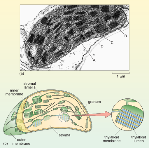
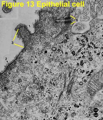
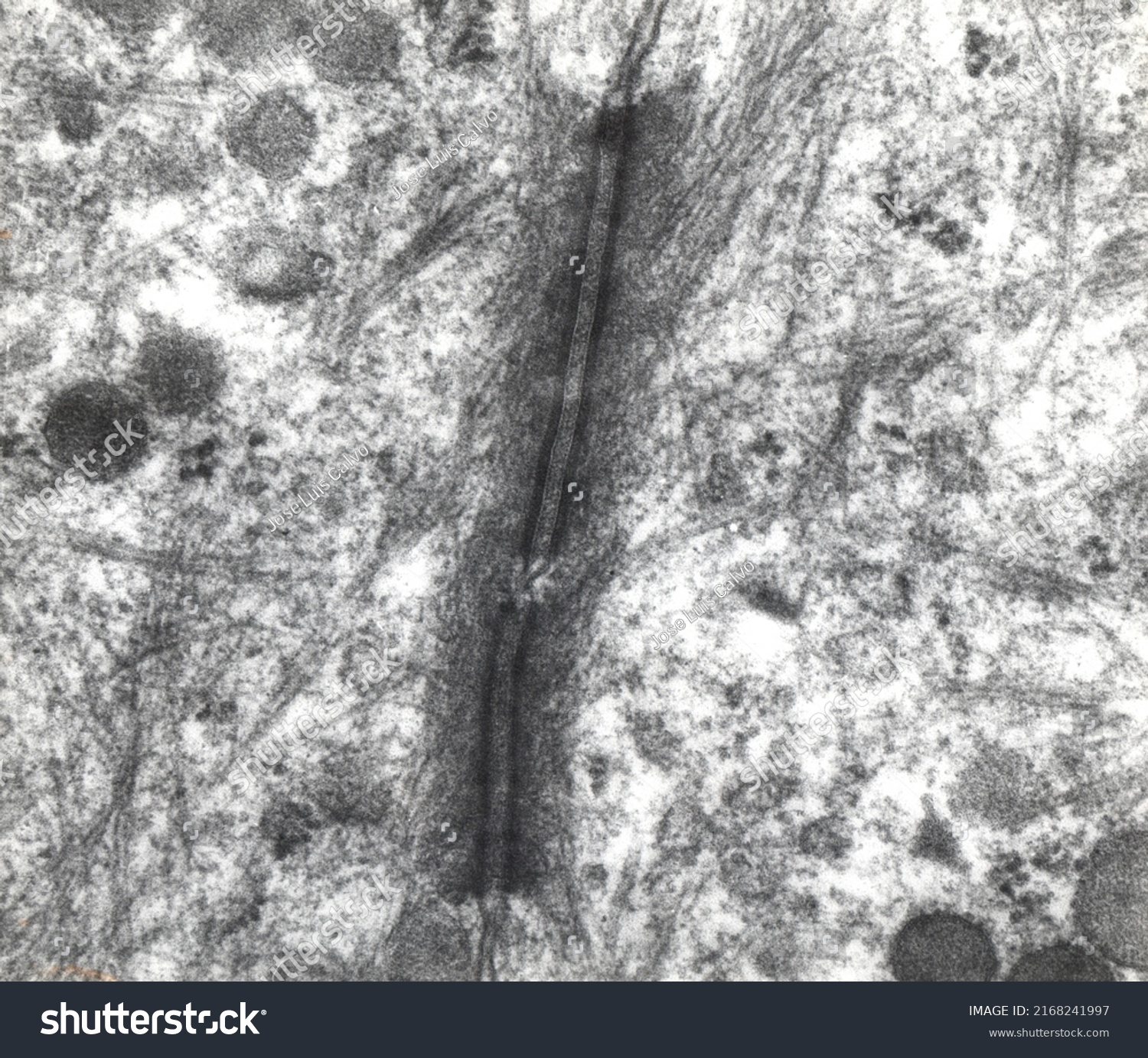
39 label the transmission electron micrograph of the cell
en.wikipedia.org › wiki › Fluorescence_microscopeFluorescence microscope - Wikipedia Fluorescence micrograph gallery A z-projection of an osteosarcoma cell, stained with phalloidin to visualise actin filaments. The image was taken on a confocal microscope, and the subsequent deconvolution was done using an experimentally derived point spread function. › cell › fulltextBacPROTACs mediate targeted protein degradation in ... - Cell Jun 03, 2022 · As seen for pArg and cyclomarin head groups, various molecules that bind to the substrate receptor of the ClpCP protease can be incorporated into a functional degrader. Using cell permeable BacPROTACs, we furthermore demonstrate that recruitment of model proteins to ClpCP leads to selective protein degradation in bacterial cells.
medlineplus.gov › ency › articleImmune response: MedlinePlus Medical Encyclopedia Lymphocytes are a type of white blood cell. There are B and T type lymphocytes. B lymphocytes become cells that produce antibodies. Antibodies attach to a specific antigen and make it easier for the immune cells to destroy the antigen. T lymphocytes attack antigens directly and help control the immune response.
Label the transmission electron micrograph of the cell
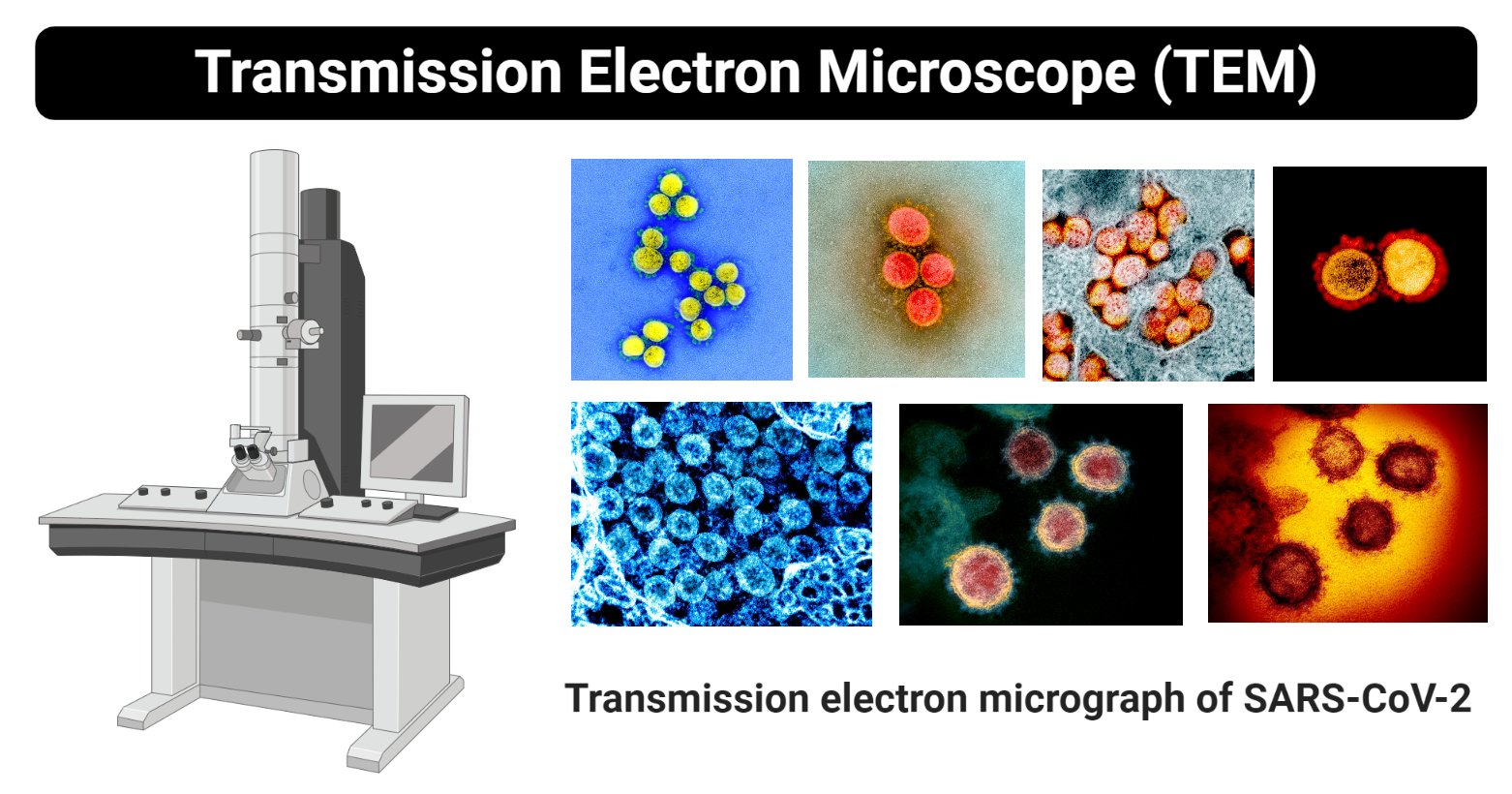
› topics › materials-scienceStacking Fault - an overview | ScienceDirect Topics Transmission electron micrograph of extended dislocations in Cu–8 at.% Ge solid-solution alloy deformed by 10%. The stacking fault energy, SFE in short, is an important parameter determining the dissociation of a dislocation, which has a significant influence on the plastic deformation behavior. › national-news › dancer-tells-ofDancer tells of excruciating pain, isolation and stigma of ... Jul 22, 2022 · A colourized transmission electron micrograph of monkeypox particles (yellow) found within an infected cell (blue), is shown in a handout photo captured at the NIAID Integrated Research Facility (IRF) in Fort Detrick, Maryland. THE CANADIAN PRESS/HO-National Institute of Allergy and Infectious Diseases **MANDATORY CREDIT** en.wikipedia.org › wiki › Electron_microscopeElectron microscope - Wikipedia An electron microscope is a microscope that uses a beam of accelerated electrons as a source of illumination. As the wavelength of an electron can be up to 100,000 times shorter than that of visible light photons, electron microscopes have a higher resolving power than light microscopes and can reveal the structure of smaller objects. A scanning
Label the transmission electron micrograph of the cell. › articles › ncomms15199Face classification using electronic synapses | Nature ... May 12, 2017 · Inset is a transmission electron microscope (TEM) image of the RRAM device. ( b ) An example of the typical continuous conductance tuning performance under an identical pulse train condition ... en.wikipedia.org › wiki › Electron_microscopeElectron microscope - Wikipedia An electron microscope is a microscope that uses a beam of accelerated electrons as a source of illumination. As the wavelength of an electron can be up to 100,000 times shorter than that of visible light photons, electron microscopes have a higher resolving power than light microscopes and can reveal the structure of smaller objects. A scanning › national-news › dancer-tells-ofDancer tells of excruciating pain, isolation and stigma of ... Jul 22, 2022 · A colourized transmission electron micrograph of monkeypox particles (yellow) found within an infected cell (blue), is shown in a handout photo captured at the NIAID Integrated Research Facility (IRF) in Fort Detrick, Maryland. THE CANADIAN PRESS/HO-National Institute of Allergy and Infectious Diseases **MANDATORY CREDIT** › topics › materials-scienceStacking Fault - an overview | ScienceDirect Topics Transmission electron micrograph of extended dislocations in Cu–8 at.% Ge solid-solution alloy deformed by 10%. The stacking fault energy, SFE in short, is an important parameter determining the dissociation of a dislocation, which has a significant influence on the plastic deformation behavior.
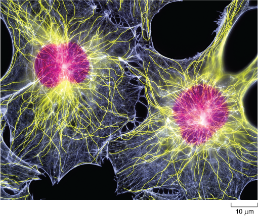
Correlative fluorescence microscopy, transmission electron ...
A tour of the cell: View as single page

2.2.3 Identify structures in electron micrographs of Ecoli
Nanomaterials | Free Full-Text | A Guide for Using ...
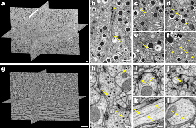
An open-access volume electron microscopy atlas of whole ...
Electron Micrographs

A genetic probe for visualizing glutamatergic synapses and ...
What is a diagram of a plant and animal cell under an ...
A tour of the cell: View as single page
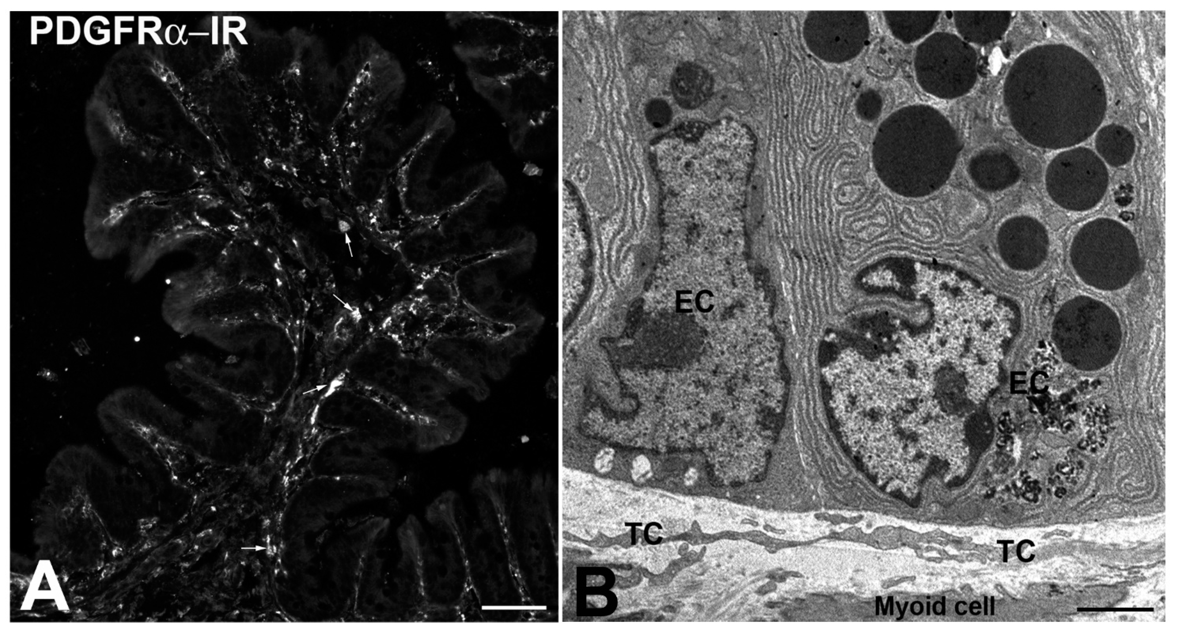
IJMS | Free Full-Text | The Telocytes: Ten Years after Their ...

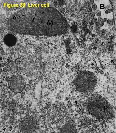
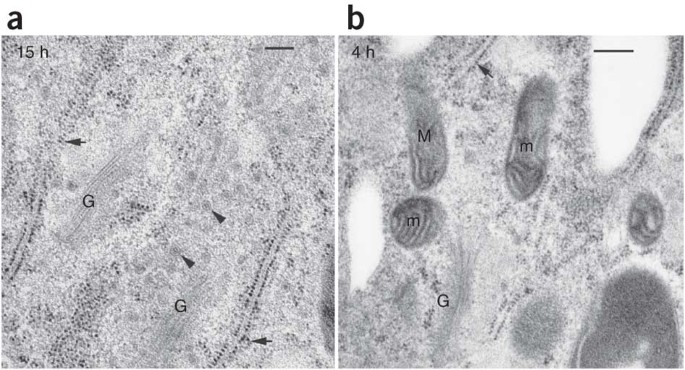
肝細胞の細胞質中のいくつかのオルガネラ(ミトコンドリア ...
Transmission electron microscope (TEM) micrograph showing the ...
Preparation of plant cells for transmission electron ...
Electron Micrographs
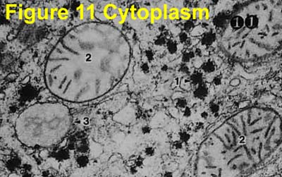
Electron Micrographs

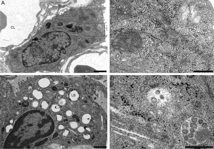
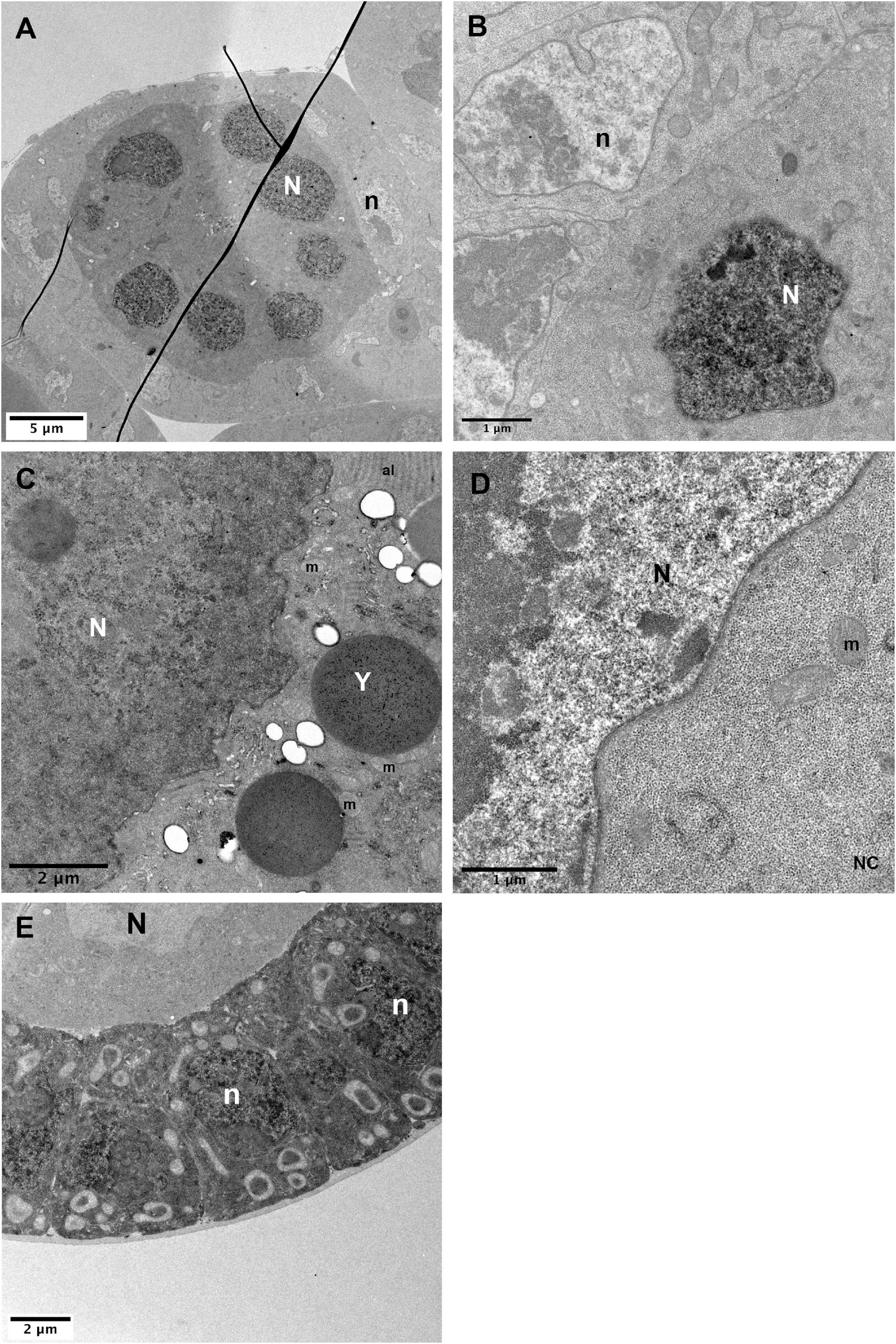
A and B) Electron micrograph of a cell labeled for/5-tubulin ...
Visualization and quantitative analysis of nanoparticles in ...

Lap Practical #1 EC Flashcards | Quizlet
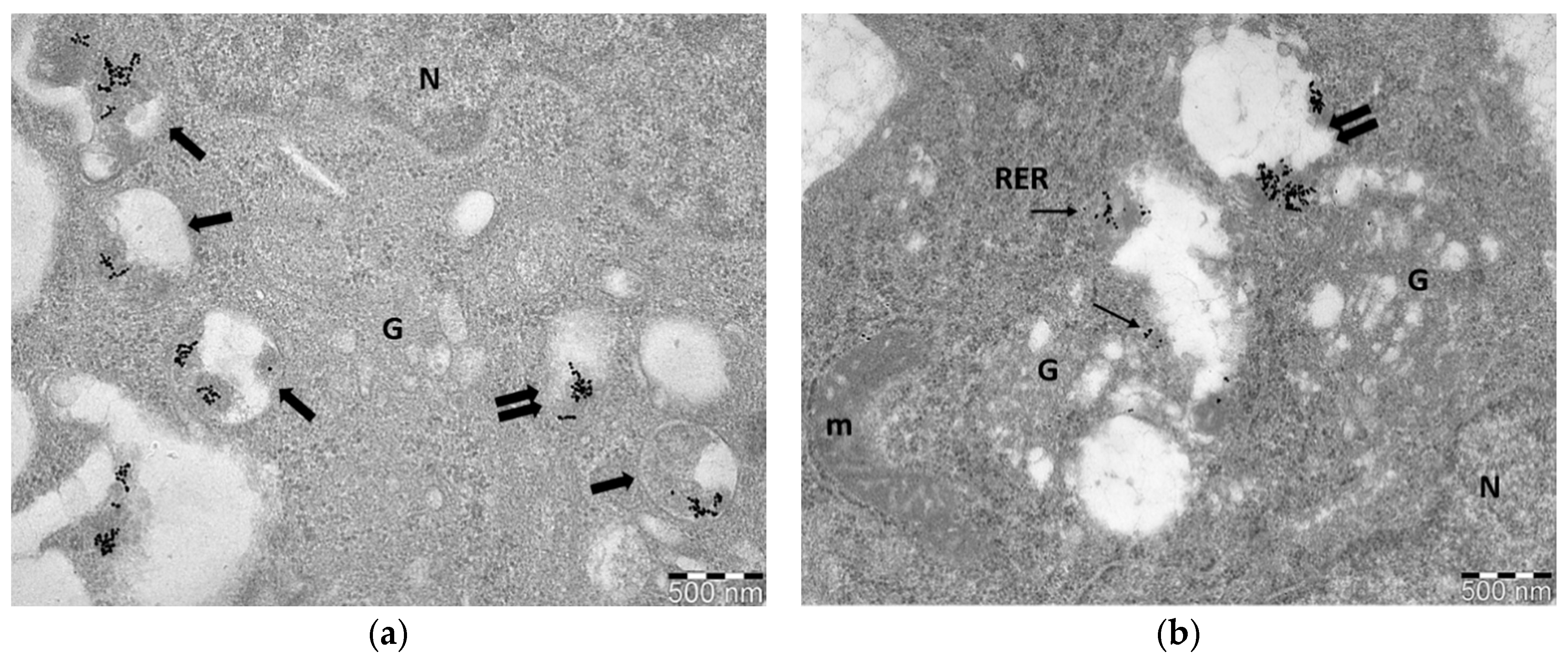
Frontiers | GFP-Tagged Protein Detection by Electron ...
ウイルス学 | アプリケーション | Leica Microsystems
Live cell immunogold labelling of RNA polymerase II ...
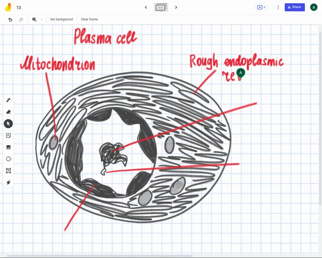
label the transmission electron ricrograph based on the hints provided mitochondnon helerochromalin plasma cell nucleus rough endoplasmic telculn nucleolus 28928

Labeling the Cell Flashcards | Quizlet

2.2.3 Identify structures in electron micrographs of Ecoli ...

Chapter 14 & 15 Flashcards Flashcards | Quizlet
High-precision targeting workflow for volume electron ...
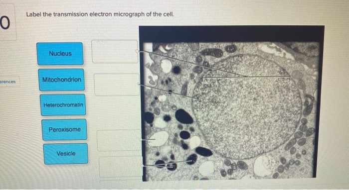
Solved Label the transmission electron micrograph of the ...

Solved Label the transmission electron micrograph based on ...
What is a diagram of a plant and animal cell under an ...
Transmission Electron Microscope (TEM)- Definition, Principle ...

Bacteria on us oh no - ppt download
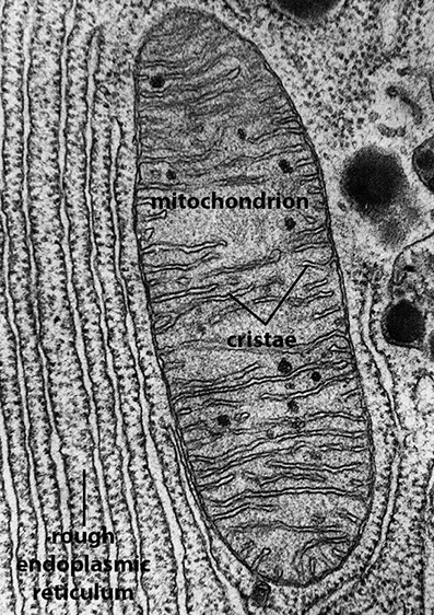
BIOL 230 Lecture Guide - Electron Micrograph of Mitochondria

Label-Free Dynamic Imaging of Chromatin in Live Cell Nuclei ...
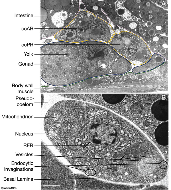
Hermaphrodite Coelomocyte System
Transmission Electron Microscope Tem Micrograph Showing写真 ...
Transmission Electron Microscopy Reveals Distinct Macrophage ...

IB Biology Skills Practice Flashcards | Quizlet

ミトコンドリア、リソソーム、rer、ゴルジ系、核(右)を示す ...
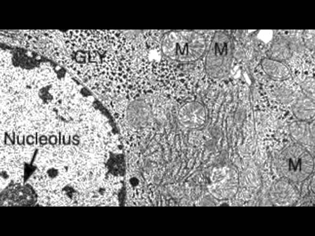
2.3.3 Identify structures from electron micrographs of liver ...
Post a Comment for "39 label the transmission electron micrograph of the cell"