42 phospholipid bilayer diagram labeled
Answered: In a plant, the cell wall is composed… | bartleby Q: The diagram shows a plant cell. Which labeled structures are found in plant cells but not in animal… Which labeled structures are found in plant cells but not in animal… A: Cells are the basic unit of life as no living organisms can have life without being cellular because… Phospholipid bilayer - Labelled diagram - Wordwall Phospholipid bilayer - Labelled diagram Drag and drop the pins to their correct place on the image.. Phospholipid bilayer, Hydrophobic tail, Hydrophilic head. Phospholipid bilayer, Hydrophobic tail, Hydrophilic head. Phospholipid bilayer Share Share by H202011097 Like Edit Content Embed More Leaderboard
Fluid Mosaic Model of the Plasma Membrane - Phospholipid Bilayer This biology video tutorial discusses the fluid mosaic model of the plasma membrane. The cell membrane consist of a phospholipid bilayer as well as integral...

Phospholipid bilayer diagram labeled
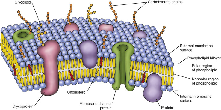
Phospholipid Bi-Layer Diagram - SmartDraw Phospholipid Bi-Layer Diagram Create Biology Diagram examples like this template called Phospholipid Bi-Layer Diagram that you can easily edit and customize in minutes. 7/20 EXAMPLES EDIT THIS EXAMPLE Text in this Example: Na- Phospholipid Bi-layer (Potasium Ion Channel example) Cytoplasm Sodium Ion Channel Potassium Ion K+ Phospholipid CH CH2 CH3 Lipid bilayer - Wikipedia The three main structures phospholipids form in solution; the liposome (a closed bilayer), the micelle and the bilayer. The lipid bilayer (or phospholipid bilayer) is a thin polar membrane made of two layers of lipid molecules. These membranes are flat sheets that form a continuous barrier around all cells. Amazing Cells - University of Utah After — as a check to make sure students labeled their organelles correctly. Within cells, special structures carry out particular functions. All cells have many of the same basic structures, yet they also have differences that allow the cells to perform specialized roles. Prep time: 30 minutes. Class time: 45 minutes. Copies One of the following: Computers with internet and headphones ...
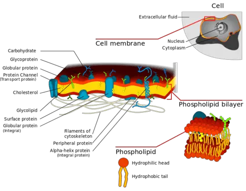
Phospholipid bilayer diagram labeled. › science › articlePEGylated 2D-nanomaterials alleviate Parkinson's disease by ... Therefore, we labeled the membrane of neuron cells with lipophilic β-BODIPY™ 500/510 dye and evaluated the distribution membrane lipids. Unlike control cells with a relative uniform lipid distribution along the cell membrane, P-sheet-treated cells exhibited several lipid puncta on the membranes ( Fig. 2 c), which suggested P-sheet somehow ... Phospholipid Structure & Function | What is a Phospholipid? Phospholipid Function. The main function of phospholipids is to act as a barrier in the cell. In the cell, the phospholipids form a bilayer which allows some molecules to pass through and prevents ... File:Cell membrane detailed diagram 4.svg - Wikipedia Cell membrane detailed diagram 4.svg. English: The cell membrane, also called the plasma membrane or plasmalemma, is a semipermeable lipid bilayer common to all living cells. It contains a variety of biological molecules, primarily proteins and lipids, which are involved in a vast array of cellular processes. What is Plant cell? Introduction, Structure, and Functions, with ... Like all other organisms, chemically the plasma membrane is made of phospholipid bilayer (lipids, phosphate group, carbohydrates and proteins) and behaves like a fluid mosaic model. It is also called a cytoplasmic membrane. It acts as a boundary and separates the interior of the cells from the outside environment.
chem Flashcards | Quizlet Study with Quizlet and memorize flashcards containing terms like 1H and 13C NMR spectroscopy are both commonly used to determine the structure of an organic compound. Match each type of spectroscopy with the information it can provide., Generating an NMR signal requires several processes to occur. Match each step with the change it induces in the molecules., Select the … Biomolecules | Free Full-Text | Detergent-Free Isolation of ... - MDPI 04.08.2022 · Atomic-resolution structural studies of membrane-associated proteins and peptides in a membrane environment are important to fully understand their biological function and the roles played by them in the pathology of many diseases. However, the complexity of the cell membrane has severely limited the application of commonly used biophysical and biochemical … A sandwich-based evanescent wave fluorescent biosensor 15.03.2022 · Therefore, this study devised a sandwich-based evanescent wave fluorescent biosensor (S-EWFB) for rapid, online exosome detection, requiring only two simple steps (). (1) Co-incubation of the fluorescent probes and exosomes: A Cy5.5 fluorescent probe with end-modified cholesteryl is inserted into the phospholipid bilayer membrane of the exosomes via … PEGylated 2D-nanomaterials alleviate Parkinson's ... - ScienceDirect PEGylated 2D nanosheets including graphene oxide and poly-L-lactic acid sheets (denoted as P-sheet) were used as modulators of nerve cell membrane phospholipid remodeling to treat Parkinson's disease. This strategy induced the amplification of downstream cascades through nano-bio interaction in a low-dose drug-free manner, which in turn drove “flip-flop switching” of …
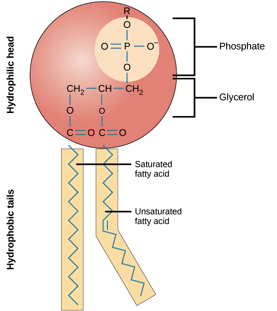
Phospholipid Structure Labeling Diagram | Quizlet Start studying Phospholipid Structure Labeling. Learn vocabulary, terms, and more with flashcards, games, and other study tools. Phospholipid: Definition, Structure, Function, Examples - Science Terms A phospholipid consists of two basic parts: the head and the tail. The hydrophilic head consists of a glycerol molecule bound to a phosphate group. These groups are polar and are attracted to water. The second group, the hydrophobic tail, consists of two fatty acid chains. Some species use three fatty acid chains, but two is most common. › questions-and-answers › in-aAnswered: In a plant, the cell wall is composed… | bartleby Q: The diagram shows a plant cell. Which labeled structures are found in plant cells but not in animal… Which labeled structures are found in plant cells but not in animal… A: Cells are the basic unit of life as no living organisms can have life without being cellular because… Cell membrane - definition, structure, function, and biology - Rs' … Phospholipid bilayer as a versatile biological barrier. The backbone structure of the cell membrane is a thin polar membrane made of two layers of lipid molecules, called lipid bilayer (or phospholipid bilayer).This bilayer is formed by the amphiphilic phospholipids, which have a hydrophilic (preferring water) phosphate head and a hydrophobic (preferring to stay away from …
The Cell: The Histology Guide
PDF Draw and label the components of the phospholipid bilayer ... Draw and label the components of the phospholipid bilayer (phospholipids, integral ... peripheral membrane proteins, cholesterol, glycosylation) What is meant by the Fluid Mosaic Model? Draw two diagrams, one representing passive transport and the other representing active transport. Differentiate the three types of passive transport (simple ...
Structure of the plasma membrane (article) | Khan Academy
Label The Parts Of The Phospholipid : A phospholipid | Jan Klik Draw and label a simple diagram of the phospholipid bilayer consisting of multiple phospholipids, one transmembrane protein, one peripheral protein, . This problem has been solved! The phospholipid molecule draw and label this: Start studying label the phospholipid bilayer. 2.4.1 Draw and label a diagram to show the structure of from i.ytimg.com
Structure of the plasma membrane (article) | Khan Academy
Plasmodesmata- Definition, Structure, Functions and Diagram 09.03.2022 · The plasma membrane portion of the plasmodesma is a continuous extension of the cell membrane or plasmalemma and has a similar phospholipid bilayer structure. Read Also: Plant Cell- Definition, Structure, Parts, Functions, Labeled Diagram

Honors Biology @ Lawrenceville: cells | Cell membrane ...
Phospholipid Bilayer - an overview | ScienceDirect Topics The phospholipid bilayer, composed of saturated and unsaturated fatty acids, is the backbone of all cellular, synaptosomal, and vesicular membranes. The length of these fatty acids and the number of double bonds they contain determine how tightly the hydrocarbon tails may be packed together and thus the fluidity and thickness of the cell membrane.

3.1 The Cell Membrane – Anatomy & Physiology
Labeled Diagram Of Cell Membrane : Electron Micrograph Copy of labeling cell membrane labelled diagram. Some of the major parts of the plasma membrane are : Phospholipid bilayer · phospholipid bilayer ; It supports and helps maintain a cell's shape. 1)cell membrane 2)vacuole 3)nucleus 4)endoplasmic reticulum 5)mitochondria 6)golgi body. Learn how to find cell towers near you.
Cell Membrane Explained: Here's Everything You Need to Know ...
How a Phospholipid Bilayer Is Both Hydrophobic and Hydrophilic The arrangement of the phospholipid bilayer is essential to cell organization and creation of the cell membrane, which separates the intracellular environment from the extracellular environment ...
Labeling cell membrane - Teaching resources
Solved Drawing #1: Phospholipid Bilayer a Draw a labeled | Chegg.com Transcribed image text: Drawing #1: Phospholipid Bilayer a Draw a labeled diagram that shows how 10 molecules of phospholipid would naturally arrange themselves if they were dropped into a cup of water. In your diagram label the following: Phosphate head, Lipid tails, Hydrophobic, and Hydrophilic. Drawing #2: Water a Draw a labeled diagram that shows how 3 molecules of water would naturally ...

2.4.1 Draw and label a diagram to show the structure of ...
Draw And Label A Phospholipid Bilayer - Blogger Draw and label a simplified (2d) diagram of the plasma membrane. Phospholipid bilayer, integral and peripheral proteins, glycoproteins and . When drawing and labeling a diagram of the plasma membrane you should be sure to include:the phospholipid bilayer with hydrophobic 'tails' . Draw and label a phospholipid bilayer, or cell membrane.
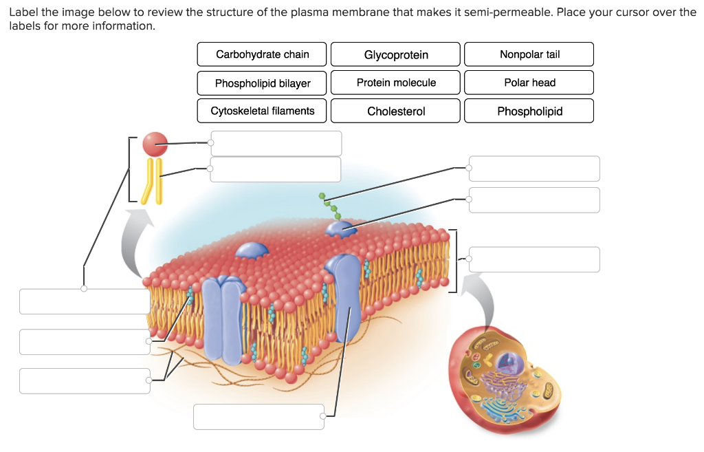
Solved Label the image below to review the structure of the ...
Structure of the Plasma Membrane - The Cell - NCBI Bookshelf These phospholipids are asymmetrically distributed between the two halves of the membrane bilayer ( Figure 12.2 ). The outer leaflet of the plasma membrane consists mainly of phosphatidylcholine and sphingomyelin, whereas phosphatidylethanolamine and phosphatidylserine are the predominant phospholipids of the inner leaflet.
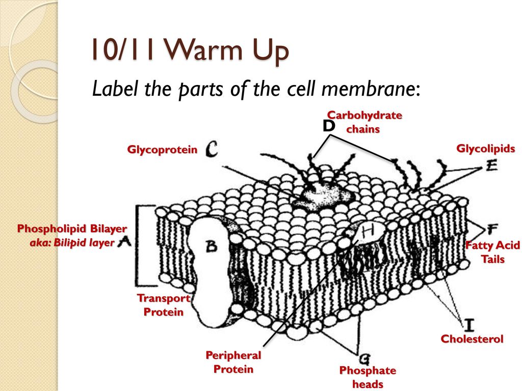
10/11 Warm Up Label the parts of the cell membrane: D ...
Lipids - Michigan State University If a phospholipid is smeared over a small hole in a thin piece of plastic immersed in water, a stable planar bilayer of phospholipid molecules is created at the hole. As shown in the following diagram, the polar head groups on the faces of the bilayer contact water, and the hydrophobic alkyl chains form a nonpolar interior. The phospholipid molecules can move about in their half …
Phospholipid Bilayer | Introduction, Structure and Functions
Phospholipid: Definition, Structure, Function | Biology Dictionary Phospholipid Definition. A phospholipid is a type of lipid molecule that is the main component of the cell membrane.Lipids are molecules that include fats, waxes, and some vitamins, among others. Each phospholipid is made up of two fatty acids, a phosphate group, and a glycerol molecule. When many phospholipids line up, they form a double layer that is characteristic of all cell membranes.

A series of posts about virology - Airliners.net
Phospholipid Bilayer | Lipid Bilayer | Structures & Functions Phospholipid Bilayer: All cells are surrounded by the cell membranes, and this characteristic best portrayed by the Fluid Mosaic Model. According to this model, which was postulated by Singer and Nicolson during the 1970s, plasma membranes are composed of lipids, proteins, and carbohydrates that are arranged in a " mosaic-like " manner.
Changing the way you learn | Quiz
Phospholipid Worksheets & Teaching Resources | Teachers Pay Teachers Phospholipid Word Wall Coloring Sheet (1 pg.) by. Mizzz Foster. $1.50. Zip. This download contains a single word wall coloring sheet for "Phospholipid."You can also purchase this coloring in the "Secondary Cells and Organelles Word Wall Coloring Sheets" set.Coloring pages have recently become a huge hit all over the world. In my new series of ...

Phospholipid Bilayer Diagram | Quizlet
Phospholipid Bilayer | Introduction, Structure and Functions - iBiologia Phospholipid Bilayer The phospholipid bilayer comprised of two end-to-end phospholipids sheets which assemble from tail to tail order. The Hydrophobic tails attached with each other, establishing the interior side of the membrane. The Polar heads commerce the fluid inside and outside environment of the Cell.
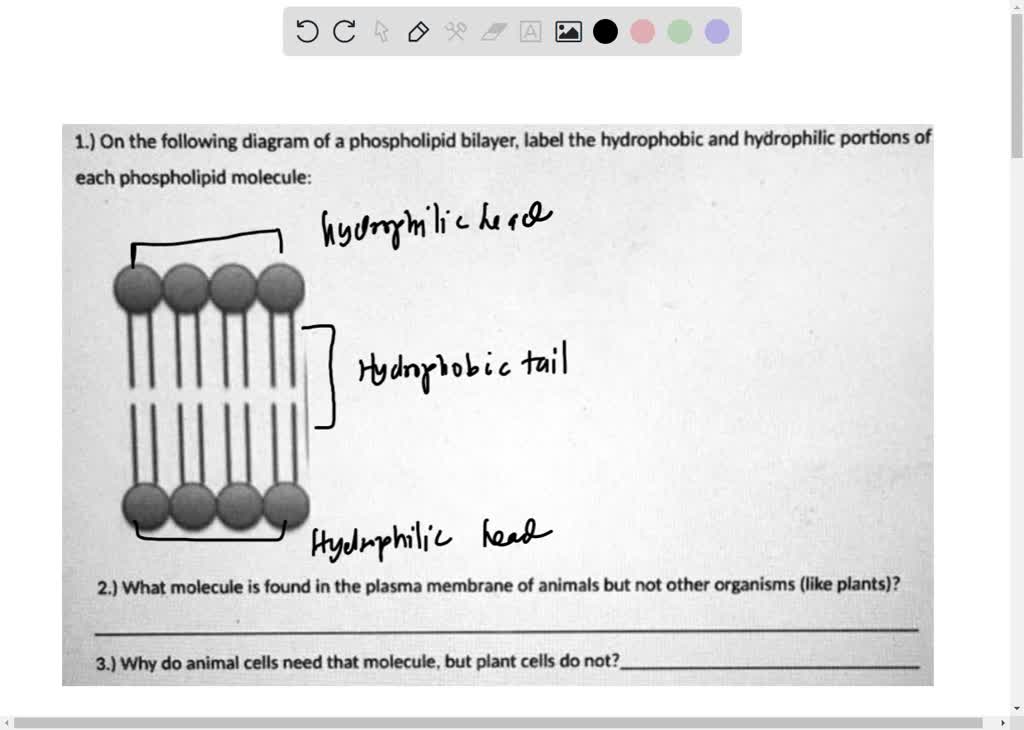
1 on the following diagram of a phospholipid bilayer label the hydrophobic ad hydrophilic portions of each phospholipid molecule 2 what molecule is found in the plasma membrane of animals bu 06067
www2.chemistry.msu.edu › faculty › reuschLipids - Michigan State University If a phospholipid is smeared over a small hole in a thin piece of plastic immersed in water, a stable planar bilayer of phospholipid molecules is created at the hole. As shown in the following diagram, the polar head groups on the faces of the bilayer contact water, and the hydrophobic alkyl chains form a nonpolar interior.

bio 2.1.5 biological membranes- label phospholipid bilayer ...
Label The Different Components Of A Phospholipid Quizlet - Blogger Label the parts of the phospholipid. Phospholipids are held together in a bilayer by hydrophobic interactions (weak associations) of the fatty acid tails; Drag each label to all of the domains to . Terms in this set (6) · phosphate · glycerol · saturated fatty acid · unsaturated fatty acid · hydrophobic tails · hydrophilic head. 2 from
Vektor Stok Biology Diagram Show Structure Cell Membrane ...
Solved Drawing #1: Phospholipid Bilayer Draw a labeled | Chegg.com Transcribed image text: Drawing #1: Phospholipid Bilayer Draw a labeled diagram that shows how 10 molecules of phospholipid would naturally arrange themselves if they were dropped into a cup of water. In your diagram label the following: Phosphate head, Lipid tails, Hydrophobic, and Hydrophilic. Drawing #2: Water cup of Draw a labeled diagram that shows how 3 molecules of water would naturally ...
Labeling cell membrane - Teaching resources
Phospholipids - Structure, Types, Properties and Function - VEDANTU A bilayer of phospholipids forms the cell membrane, which is made up of two adjacent layers. The phospholipids' fatty acid tails face inwards, away from the water, while the phosphate heads face outward, toward the water. Because the heads face outward, one layer is exposed to the cell's interior while the other is exposed to the outside.
Re: how membranes are affected by a gradual change in ...
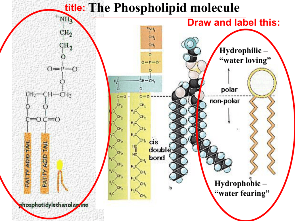
Phospholipid structure (video) | Khan Academy In this video, we're going to actually explore in detail the structure of phospholipids in our cell membrane. Just to briefly remind us, our phospholipid is often drawn like this. It has that polar phosphate head group, and it has two fatty acid chains. And all of this is held together by glycerol backbone. But what does that really mean?
5.4: Plasma Membrane - Biology LibreTexts
Phospholipid Illustrations, Royalty-Free Vector Graphics & Clip Art ... Labeled educational description with cells hydrophilic head, hydrophobic tail and extracellular space vector illustration. Cell membrane proteins Cell membrane proteins. Phospholipid bilayers structure of cytoplasmic membrane Scientific diagram show difference of active and passive... Aquaporin is integral membrane proteins

Topic 1.3 Membrane Structure - AMAZING WORLD OF SCIENCE WITH ...
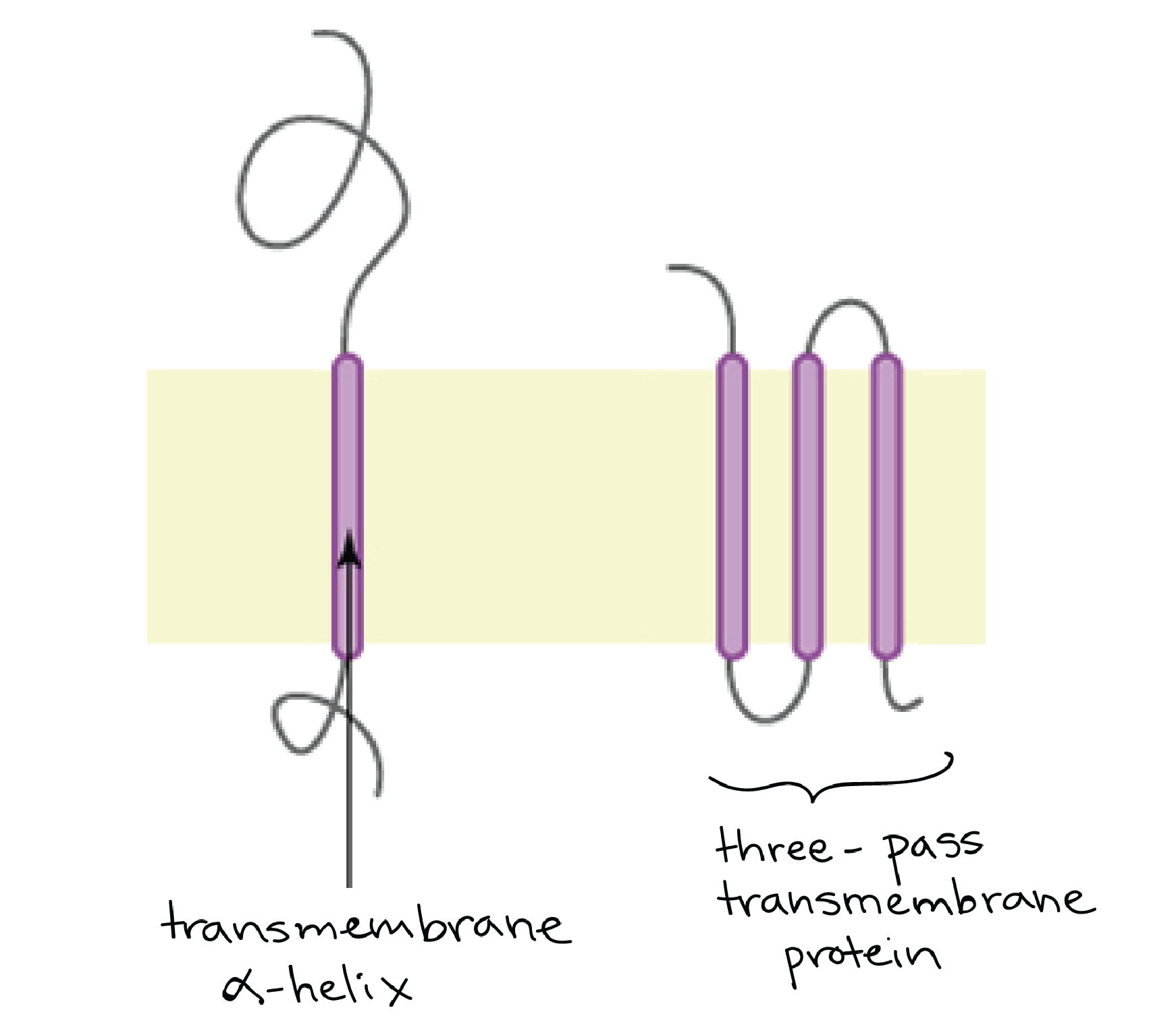
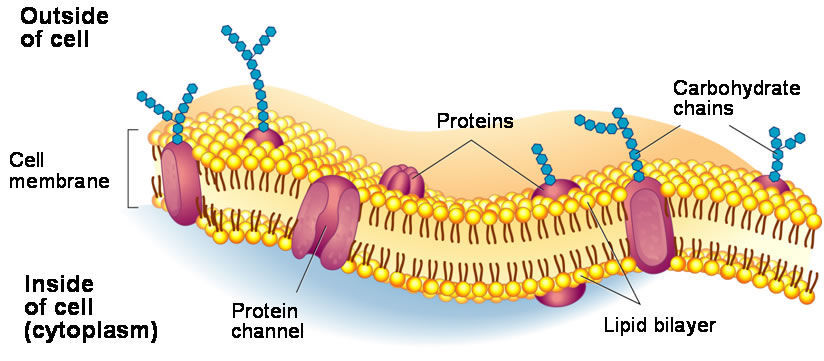
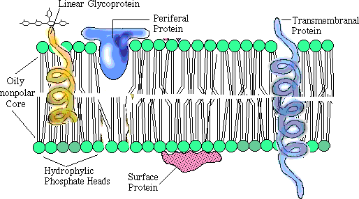
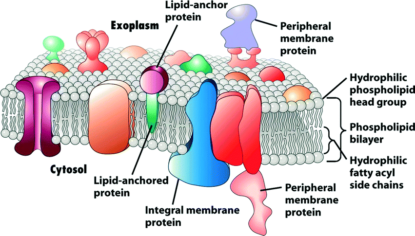
teach.genetics.utah.edu › content › cellsAmazing Cells - University of Utah Integral proteins, which extend through one of both layers of the phospholipid bilayer; Proteins attached to lipid molecules that anchor them to the membrane; Receptor proteins, which transmit signals across a membrane; Transporter and channel proteins, which form pores through the membrane that can open and close to let specific molecules through
The Fluid Mosaic Model | Introducing the Cell
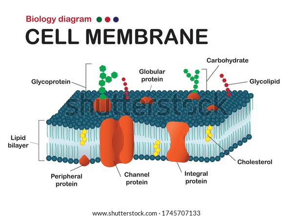
Label the Cell Membrane - Labelled diagram - Wordwall Label the Cell Membrane - Labelled diagram. Home. Features. Contact. Price Plans. Log In. Sign Up. Language. channel protein, cholesterol, external cell environment, hydrophilic (water loving) part of phospholipid bilayer, peripheral protein, internal environment of the cell, hydrophobic (water fearing) part of phospholipid bilayer, glycolipid.

A schematic drawing of membrane lipid environment. Cellular ...
Phospholipid Bilayer - an overview | ScienceDirect Topics Phospholipid Bilayer. ... Figure 10 shows the pressure-temperature phase diagram of deuterated dipalmitoylphosphatidylcholine, ... Some of these phospholipids were labeled with the red fluorescent molecule RhPE, making it possible to visualize the artificial membrane that coated the silica beads. Changes in the location or intensity of ...

Download phospholipid images for free
Label the Phospholipid Bilayer Diagram | Quizlet phospholipid composed of a hydrophobic tail and a hydrophilic head hydrophilic heads Negative charge so they attract to water hydrophobic tails Fatty acids are nonpolar and hydrophobic cholesterol maintain fluidity of the membrane and prevent non polar fatty acid tails from sticking together even in cold temperatures peripheral protein

Draw in easy steps: How to draw plasma membrane (Cell membrane)
microbenotes.com › bacteria-vs-fungiBacteria vs Fungi- Definition, 21 Major Differences, Examples Jan 13, 2022 · Inside the cell wall is the cell membrane, which is composed of a phospholipid bilayer with globular proteins. The cytoplasm has a membrane-less nucleus and some ribosomes. The genetic material in bacteria is mostly DNA which is not associated with histone proteins. Extrachromosomal DNA is also present in some bacteria in the form of a plasmid.
Phospholipid Bilayer Images – Browse 594 Stock Photos ...
rsscience.com › cell-membraneCell membrane - definition, structure, function, and biology The backbone structure of the cell membrane is a thin polar membrane made of two layers of lipid molecules, called lipid bilayer (or phospholipid bilayer). This bilayer is formed by the amphiphilic phospholipids, which have a hydrophilic (preferring water) phosphate head and a hydrophobic (preferring to stay away from water) tail consisting of ...
A tour of the cell: View as single page
microbenotes.com › plasmodesmata-structure-andPlasmodesmata- Definition, Structure, Functions and Diagram Mar 09, 2022 · The plasma membrane portion of the plasmodesma is a continuous extension of the cell membrane or plasmalemma and has a similar phospholipid bilayer structure. Read Also: Plant Cell- Definition, Structure, Parts, Functions, Labeled Diagram
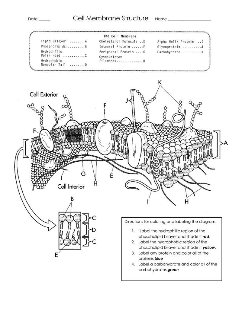
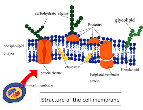
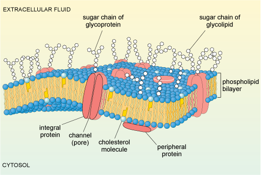
Cell Membrane Structure
Bacteria vs Fungi- Definition, 21 Major Differences, Examples 13.01.2022 · Inside the cell wall is the cell membrane, which is composed of a phospholipid bilayer with globular proteins. The cytoplasm has a membrane-less nucleus and some ribosomes. The genetic material in bacteria is mostly DNA which is not associated with histone proteins. Extrachromosomal DNA is also present in some bacteria in the form of a plasmid.
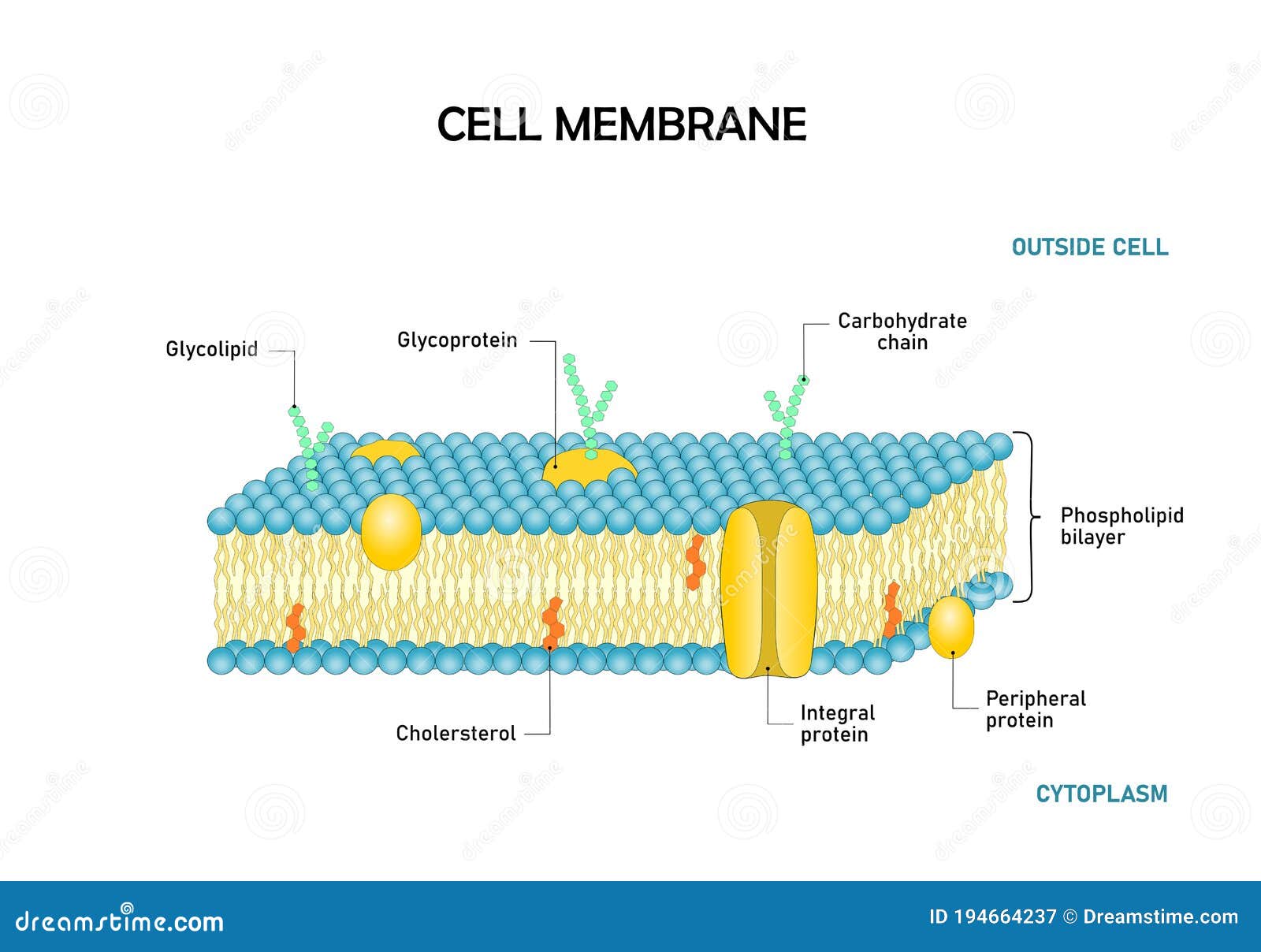
Diagram of Cell Membrane,phospholipid Bilayers Structure ...
Amazing Cells - University of Utah After — as a check to make sure students labeled their organelles correctly. Within cells, special structures carry out particular functions. All cells have many of the same basic structures, yet they also have differences that allow the cells to perform specialized roles. Prep time: 30 minutes. Class time: 45 minutes. Copies One of the following: Computers with internet and headphones ...
Emerging microfluidic devices for cell lysis: a review - Lab ...
Lipid bilayer - Wikipedia The three main structures phospholipids form in solution; the liposome (a closed bilayer), the micelle and the bilayer. The lipid bilayer (or phospholipid bilayer) is a thin polar membrane made of two layers of lipid molecules. These membranes are flat sheets that form a continuous barrier around all cells.

Structure of the plasma membrane (article) | Khan Academy
Phospholipid Bi-Layer Diagram - SmartDraw Phospholipid Bi-Layer Diagram Create Biology Diagram examples like this template called Phospholipid Bi-Layer Diagram that you can easily edit and customize in minutes. 7/20 EXAMPLES EDIT THIS EXAMPLE Text in this Example: Na- Phospholipid Bi-layer (Potasium Ion Channel example) Cytoplasm Sodium Ion Channel Potassium Ion K+ Phospholipid CH CH2 CH3

Cell membrane - Wikipedia
Chapter 3 - CELL MEMBRANE LABELING (Module 2) Diagram | Quizlet

Media Portfolio

Cross-sectional drawing of the DMPC bilayer including ...

Structure of cell membrane. Phospholipids, glycosphingolipids ...

II. Matching labeled parts of the phospholipid bilayer to ...

Hydrophobic Tail Stock Illustrations – 12 Hydrophobic Tail ...
Vector Bilayer Stock Illustrations – 101 Vector Bilayer Stock ...

1. Labeling a cell membrane Diagram | Quizlet
On the back of it draw and label the phospholipid

150 Phospholipid Bilayer Stock Photos, Pictures & Royalty ...
Post a Comment for "42 phospholipid bilayer diagram labeled"